Application of a silver and platinum nanocluster in tumor-targeted imaging

A tumor-targeted imaging and platinum nanotechnology, which is applied in the field of medical imaging, can solve the problems of no silver and platinum nanoclusters, and achieve the effects of small particle size, good absorption and good permeability

Active Publication Date: 2016-04-13
SOUTHEAST UNIV
View PDF4 Cites 0 Cited by
  • Summary
  • Abstract
  • Description
  • Claims
  • Application Information

AI Technical Summary

Problems solved by technology

[0004] Chinese patent CN102735752A discloses the application of gold nanoclusters in tumor-targeted in vivo imaging. At present, there is no literature on the application of silver and platinum nanoclusters in tumor-targeted imaging.

Method used


Image

  • Application of a silver and platinum nanocluster in tumor-targeted imaging
  • Application of a silver and platinum nanocluster in tumor-targeted imaging
  • Application of a silver and platinum nanocluster in tumor-targeted imaging

Examples


Embodiment 1

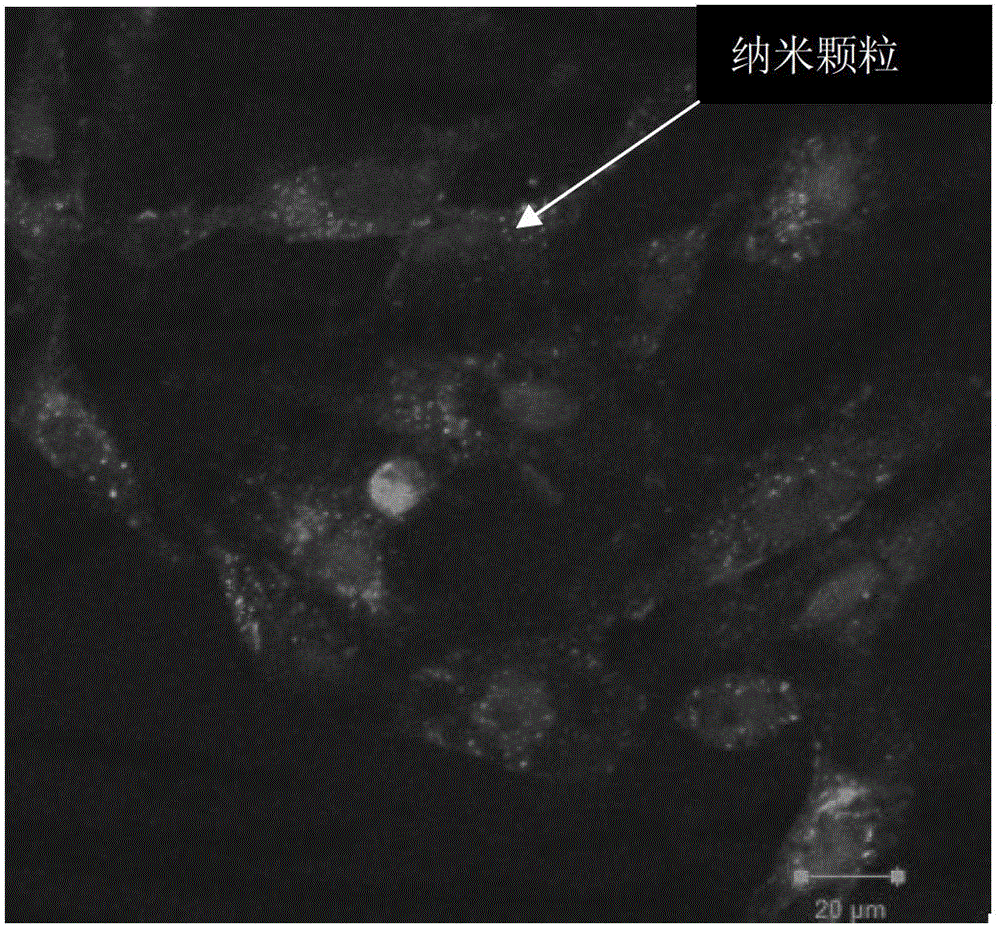
[0026] Liver cancer cells (HepG2) were selected as the research object, and the experimental group divided the liver cancer cells (HepG2) in the logarithmic growth phase at a ratio of 1.6×10 5 The density of cells / well was inoculated in 6-well plates, and after 24 hours of culture, 1 μmol / L solution of glutathione-silver ammonia that had been sterilized and diluted with fresh sterile DMEM medium was added.

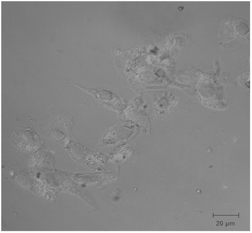
[0027] In the control group, the liver cancer cells (HepG2) in the culture medium were divided into 1.6×10 5 Cells / well were seeded in 6-well plates and cultured for 24 h. After the incubation time was terminated, phosphate buffer solution (PBS, pH=7.4) was added to each well of the experimental group and the control group to wash 2-3 times. Then observe and detect with a confocal microscope. The experimental results of the experimental group are as follows: Figure 1a As shown, the aggregation of silver nanoparticles can be observed, and the experimental results of the ...

Embodiment 2

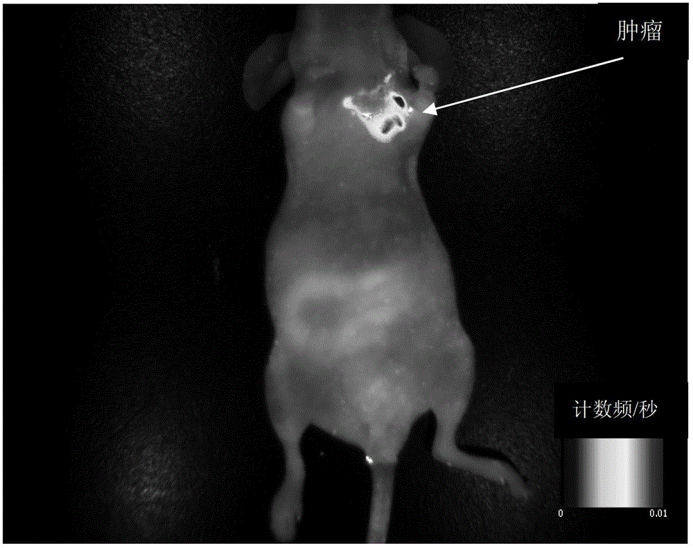
[0029] Construct the breast cancer model subcutaneously inoculated in nude mice. After the breast cancer cells are cultured for 24 hours, add 0.1 μmol / L glutathione silver ammonia solution that has been sterilized and diluted with fresh sterile DMEM medium, and incubate in the cell culture incubator Silver nanoclusters were prepared in 24 hours, and the solution containing silver nanoclusters was ultrasonically dispersed and injected into nude mice through tail vein injection. After 24 hours, in vivo fluorescence imaging was performed on nude mice. Experiments have shown that silver nanoclusters can achieve targeted imaging of tumor sites. Experimental results such as figure 2 shown.

Embodiment 3

[0031] The orthotopic transplantation model of liver cancer in nude mice was constructed. After the liver cancer cells were cultured for 24 hours, 50 μmol / L cis-diaminodibromoplatin solution, which had been sterilized and diluted with fresh sterile DMEM medium, was added and incubated in the cell culture box for 48 hours. Platinum nanoclusters were prepared in 1 hour, and the solution containing platinum nanoclusters was ultrasonically dispersed and injected into nude mice by local subcutaneous injection. After 24 hours, nuclear magnetic imaging was performed on nude mice and the imaging results were qualitatively and quantitatively analyzed.


PUM


Abstract

An application of a silver and platinum nano-cluster in tumor targeted imaging comprises the steps of: establishing a nude mouse tumor model; locally injecting an ultrasonically dispersed silver or platinum nano-cluster into a nude mouse, or directly injecting a silver or platinum containing solution of 0.1 to 100 μmol / L into a nude mouse; and in 24 hours after injection, performing medical imaging on the tumor part of the nude mouse by using a medical imaging method, and performing qualitative and quantitative analysis on the imaging result.

Description

technical field [0001] The invention relates to the application of silver and platinum nano-clusters as tumor imaging agents in tumor targeting imaging, and the invention is applicable to the field of medical imaging. Background technique [0002] In vivo bioimaging technology is a powerful means to detect molecular and cellular events in experimental animals. It is being more and more widely used in the field of medical and biological research. It has extremely high detection sensitivity and low experimental cost. However, the technology still has many limitations, especially the tissue autofluorescence and Poor tissue permeability under broken wave excitation light. [0003] In order to overcome these difficulties. Scientists have studied a series of inorganic luminescent nanomaterials. Some inorganic nanomaterials, such as silicon nanoparticles, are widely used in biological analysis and imaging due to their excellent properties, but there are still some problems, the m...

Claims


Application Information

Patent Timeline
Patent Type & Authority Patents(China)
IPC IPC(8): A61K49/00A61K49/06A61K49/04A61K49/22
CPCA61K49/0002A61K49/0065A61K49/0414A61K49/1818A61K49/225
Inventor 王雪梅高生平吴长宇李水红叶静
Owner SOUTHEAST UNIV
Why Eureka
Try Eureka