Gene engineering antibody for kitasamycin residue detection, and preparation method and application thereof

A technology of genetically engineered antibodies and kitasamycin, which is applied in the directions of genetic engineering, botany equipment and methods, biochemical equipment and methods, etc., can solve the problems of unobtained patent documents, achieve easy production, high detection accuracy, reduce The effect of fees

Active Publication Date: 2013-09-11
HUAZHONG AGRICULTURAL UNIVERSITY
View PDF2 Cites 3 Cited by
  • Summary
  • Abstract
  • Description
  • Claims
  • Application Information

AI Technical Summary

Problems solved by technology

According to the results of literature search, no patent literature related to the subject of this research was obtained

Method used


Image

  • Gene engineering antibody for kitasamycin residue detection, and preparation method and application thereof
  • Gene engineering antibody for kitasamycin residue detection, and preparation method and application thereof
  • Gene engineering antibody for kitasamycin residue detection, and preparation method and application thereof

Examples


Embodiment 1

[0025] Example 1: KA / 2A9 cell line phage antibody library construction

[0026] Take the monoclonal hybridoma cell line KA / 2A9 preserved in liquid nitrogen (the hybridoma cell line KA / 2A9 is preserved in the China Center for Type Culture Collection, and its preservation number is CCTCC NO: C201184), and quickly thaw it in a water bath at 40°C. Dissolve 10ml RPMI-1640, centrifuge the cells horizontally at 1000rpm, discard the supernatant, pipette 1ml Trizol (Invitrogen, USA) into a centrifuge tube, invert up and down to mix well, and let stand at room temperature (20-25°C, the same below) for 5min. Add 400 μL of chloroform, shake vigorously for 15 s, and place at room temperature for 10 min. Centrifuge at 12000r / min at 4°C for 10min, and three layers can be seen at this time. Carefully take 600 μL of the supernatant, transfer it to a new EP tube, and add 600 μL of pre-cooled isopropanol. Inverted to mix, room temperature for 10min. Centrifuge at 12000r / min at 4°C for 10min. ...

Embodiment 2

[0044] Example 2: Specific antibody screening

[0045] Take 200 μl of the cryopreserved library and add it to a 250mL Erlenmeyer flask, add 50mL 2×YT-AG liquid medium (100mg / L and 2% (mass percentage) glucose), and culture at 37°C and 250r / min until OD600=0.8. Take 2×10¹⁰ M13K07 helper phage (NEB, USA) and add it to the bacterial solution. Incubate at 37°C, 250r / min for 1h. Centrifuge at 1000g for 10min, and carefully aspirate the supernatant. The samples were resuspended in 20 mL 2×YT-AK (100, 50) liquid medium, and cultured overnight at 30°C and 250 r / min. Centrifuge at 1000g for 20min. The supernatant was transferred to a 100 mL centrifuge tube, precipitated with 50% PEG, and left to stand on ice for 1 h. Centrifuge at 10,000 r / min for 10 min at 4°C, and dissolve the precipitate in 3 mL of phosphate buffer.

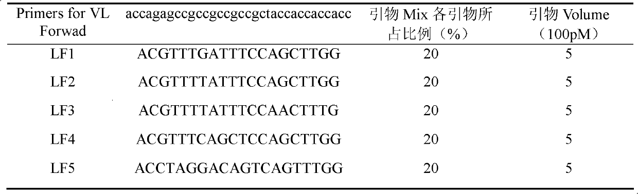
[0046] Take a sterile immune test tube, add 3 mL of coating solution, and add 30 μl of 1 mg / mL kitasarmycin coating agent according to the final coating concentra...

Embodiment 3

[0052] Example 3: Soluble Expression of Single Chain Antibody

[0053] Pick a single colony on the plate and put it into 5mL 2×YT-AG liquid medium, and culture overnight at 30°C and 250r / min. The overnight culture was inoculated with 2×YT-AG liquid medium at a ratio of 1:10, cultured at 30° C. for 1 hour at 250 r / min, and centrifuged at 1500 g for 10 minutes. The precipitate was resuspended in 50mL 2×YT-AI liquid medium, and the expression was induced at 30°C and 250r / min for 6-8h. Centrifuge the bacterial solution at 1500 g for 20 minutes, resuspend the pellet in 2% bacterial solution volume TES, add 3% volume 1 / 5×TES, mix well, and ice-bath for 40 minutes. Centrifuge at 12000r / min for 10min, and the supernatant is the periplasmic cavity extract. After aliquoting in small volumes, store at -20°C.

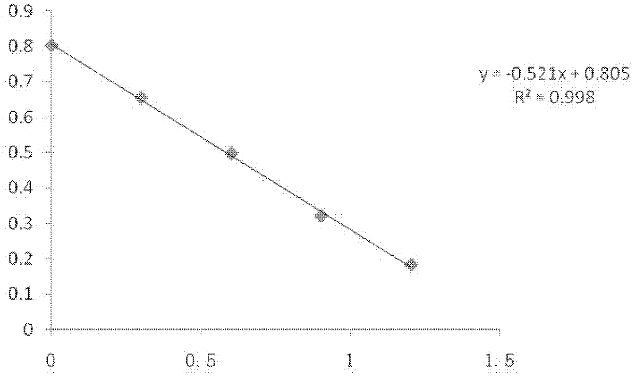
[0054] The culture supernatant and the periplasmic cavity extract were taken respectively for potency ELISA determination. The periplasmic lumen extract and the culture superna...


PUM


Abstract

The invention discloses a gene engineering antibody for kitasamycin residue detection, and a preparation method and application thereof. The preparation method comprises the following steps: A. taking hybridoma cell strain KA / 2A9 preserved in liquid nitrogen, extracting splenocyte total RNA (ribonucleic acid) to obtain mRNA (messenger ribonucleic acid), and carrying out reverse transcription to obtain cDNA (complementary deoxyribonucleic acid); B. amplifying to obtain heavy chain variable region and light chain variable region genes; by using heavy chain and light chain antibody segments as primers, connecting the antibody heavy chain with the light chain by using a polypeptide chain; C. connecting digestion scFv into E.coliTG1, adding M13K07, and culturing to obtain a bacteriophage antibody library of kitasamycin-specific antibodies; D. adding the bacteriophage into E.coliHB2151 in a logarithmic growth phase, and carrying out IPTG (isopropyl-beta-D-thiogalactopyranoside) induction to obtain a specific bacteriophage antibody; and E. sequencing the verified Escherichia coli expression strain to obtain the single chain antibody amino acid and nucleotide sequences. The method is easy to implement, simple to operate and convenient for mass production of gene engineering antibodies. The invention is widely used for detecting kitasamycin residues in animal edible tissues, and provides residue level information of kitasamycin in animal edible tissues.

Description

technical field [0001] The invention belongs to the technical field of veterinary drug residue analysis and genetic engineering antibodies, and specifically relates to a genetic engineering antibody capable of detecting kitasamycin residues, and also relates to a preparation method of a genetic engineering antibody for kitasarmycin residue detection, and also relates to the kitasarmycin residue detection method. Application in genetically engineered antibodies for detection of protein residues. Background technique [0002] Kitamycin is a macrolide antibiotic widely used in pig and poultry production. Kitamycin can cause adverse reactions in humans, mainly including mild gastrointestinal reactions such as diarrhea, nausea, vomiting, allergies, liver toxicity, tinnitus, hearing impairment and cardiotoxicity. Kitamycin residues can cause bacterial resistance in the human body and directly affect the treatment of human diseases. Bacterial drug resistance experiments in clinic...

Claims


Application Information

Patent Timeline
IPC IPC(8): C07K16/44C12N15/13G01N33/543
Inventor 袁宗辉王小清彭大鹏王玉莲黄玲利陈冬梅陶燕飞戴梦红刘振利谢长清蒋能辉
Owner HUAZHONG AGRICULTURAL UNIVERSITY
Why Eureka
Try Eureka