Green fluorescent protein radioimmunoassay kit and its preparation method and detection method

A green fluorescent protein and radioimmunoassay technology, applied in the field of biotechnology detection, can solve the problems of limited detection sensitivity and color development accuracy, difficult to control color development time, unfavorable for accurate detection, etc., and achieves easy operation and reduces non-specific adsorption. , the effect of saving antibodies

Inactive Publication Date: 2015-09-16
INST OF ANIMAL SCI OF CHINESE ACAD OF AGRI SCI
View PDF5 Cites 0 Cited by
  • Summary
  • Abstract
  • Description
  • Claims
  • Application Information

AI Technical Summary

Problems solved by technology

[0005] Quantitative detection of GFP residues by ELISA method, the color development time is not easy to control, and the detection sensitivity is limited by the accuracy of the color development of the immune reaction
For a small amount of residues, the detection rate is low, which is not conducive to accurate detection

Method used


Image

  • Green fluorescent protein radioimmunoassay kit and its preparation method and detection method
  • Green fluorescent protein radioimmunoassay kit and its preparation method and detection method
  • Green fluorescent protein radioimmunoassay kit and its preparation method and detection method

Examples


Embodiment 1

[0054] The assembly of embodiment 1 kit

[0055] 1. Buffer A: 0.1M phosphate buffer (PBS) pH7.2; 1 bottle (liquid), 20ml, shake well and use directly.

[0056] 2. Buffer B: 1% bovine serum albumin, 0.1% cold sea fish skin glue, 0.05% sodium azide, 0.1M phosphate buffer (PBS) pH7.2; 1 bottle (colorless, liquid), 30ml, shake Use directly after mixing.

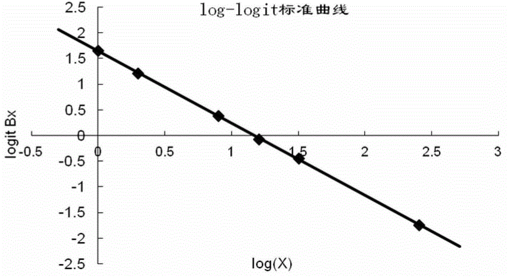
[0057]3. GFP standard product: purchased from Vector Company, 1 tube (green, liquid, 1.024ng / μl). Draw 1 μl before use, add 2ml of buffer A to dissolve, the concentration is 512pg / ml, as S10, take another 9 test tubes numbered S9-S1, add 1ml of buffer A to each, take 1ml from S10, add to S9, and serially dilute To S1, the concentration is 1, 2, 4, 8, 16, 32, 64, 128, 256 pg / ml.

[0058] 4. GFP polyclonal antibody: purchased from chemicon company, 1 bottle (colorless, liquid), absorbed before use, dissolved in buffer B, diluted 1:4000.

[0059] 5. 125 I-GFP: 1 vial (white, freeze-dried powder), dissolved in 15ml of buffer B. ...

Embodiment 2

[0061] Example 2 Determination of GFP content in blood samples of transgenic animals (cattle, pigs, etc.)

[0062] 1. Serum sample collection: Take a disposable procoagulant serum blood collection tube (BD yellow cap, colloid separation method) and a bidirectional blood collection needle (BD) to quickly puncture the jugular vein to obtain 5ml of blood, immediately gently mix it upside down 5 times, and centrifuge at 4000rpm at 4°C , 5min, separate the serum, and store it at 4°C.

[0063] 2. Measurement method

[0064] In this experiment, the balance method is used, and the operation is carried out according to the following steps:

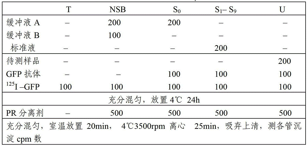
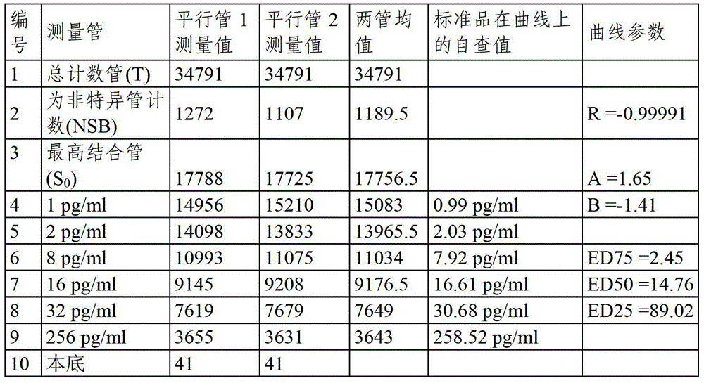
[0065] 1) Numbering on polystyrene test tubes, including main tube (T tube), non-specific tube (NSB tube), zero-binding tube (S 0 tube), standard tube (S 1 -S 9 tube), sample tube (U tube), and the above test tubes must be labeled with double tubes.

[0066] 2) To S 1 -S 9 Add 200μl standard, 100μl GFP polyclonal antibody and 100μl 125 I–GF...


PUM

PropertyMeasurementUnit
Sensitivityaaaaaaaaaa

Abstract

The invention provides a green fluorescent protein radio-immunity kit as well as a preparation method and a detection method thereof. The kit comprises radionuclide-marked green fluorescent protein, green fluorescent protein polyclonal antibodies, a buffer liquid A, a buffer liquid B, a PR (pyrogallol red) separating agent and a green fluorescent protein standard product. By utilizing the kit provided by the invention, GFR (Green Fluorescent Protein) residues in blood, organs and tissue protein solution of animals can be accurately and sensitively detected, and the lowest detection limit is 1 pg / ml. The kit has the advantages that the use concentrations of primary antibodies are low, a preprocessing process of a sample is simple and consumes little time, a large number of samples can be simultaneously detected, the experiment efficiency is high, and the sample detection cost is far less than that of a traditional instrument detection method, and the kit has very important practical significances on monitoring techniques for solving the green fluorescent protein residues of the mass samples.

Description

technical field [0001] The invention relates to the field of biotechnology detection, in particular to a green fluorescent protein radioimmunoassay kit, a preparation method and a detection method thereof. Background technique [0002] Green fluorescent protein (GFP) is a marker protein widely used in transgenic animal production. It was discovered in 1962 in a jellyfish named Aequorea victoria. The protein produced by its gene, when excited by light in the blue wavelength range, will absorb part of the energy of blue light and emit green fluorescence. This property has been exploited for in vivo biomarkers to perform real-time microscopic observation of biological samples under blue light illumination. [0003] Green fluorescent protein is a protein of lower animals. In the production of transgenic large livestock, green fluorescent protein protein is an exogenous protein. Green fluorescent protein may remain in the animal's internal organs and various organs and tissues ...

Claims


Application Information

Patent Timeline
Patent Type & Authority Patents(China)
IPC IPC(8): G01N33/68
Inventor 刘岩朱化彬
Owner INST OF ANIMAL SCI OF CHINESE ACAD OF AGRI SCI
Why Eureka
Try Eureka