Rapid neuron staining method based on Golgi silver staining method

A staining method and neuron technology, applied in the field of neuropathological tissue staining, can solve the problems of affecting the experimental process, the staining effect is not good, and the process is time-consuming, so as to simplify the traditional experimental steps, overcome the difficulty of slice operation, and overcome the long-term operation. Effect

CN103471898AInactive Publication Date: 2013-12-25HEFEI UNIV OF TECH
2 Cites 6 Cited by

Patent Information

Authority / Receiving Office
CN · China
Current Assignee / Owner
Publication Date
2013-12-25
Estimated Expiration
Not applicable · inactive patent

Smart Images

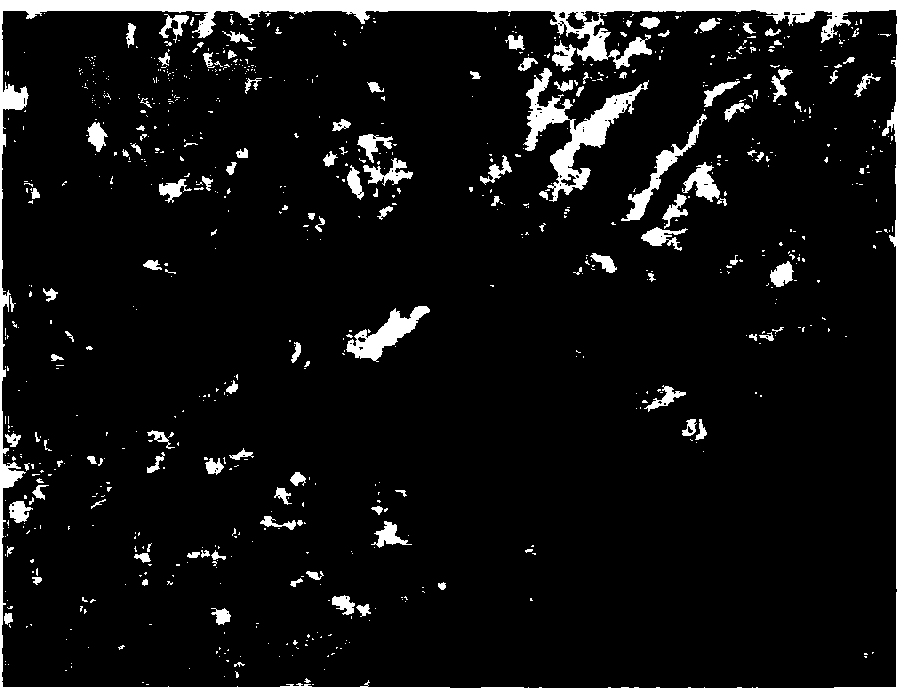
  • Figure 1
    Figure 1
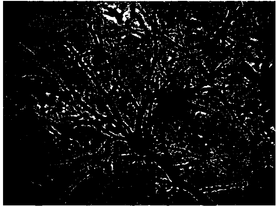
  • Figure 2
    Figure 2
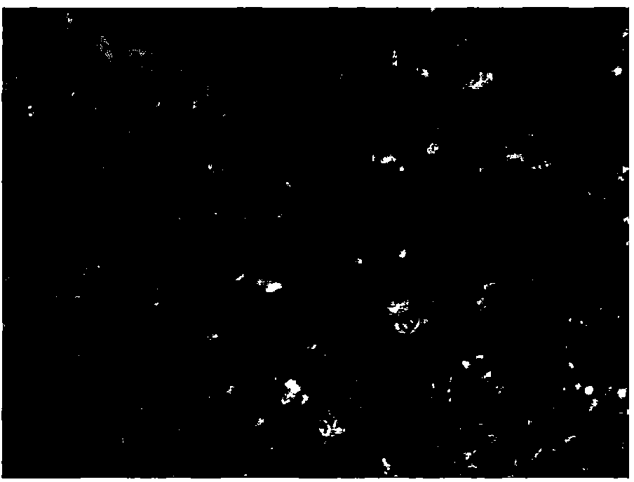
  • Figure 3
    Figure 3
Patent Text Reader

Abstract

The invention discloses a rapid neuron staining method based on a Golgi silver staining method. The rapid neuron staining method based on the Golgi silver staining method is characterized in that according to mass volume ratio concentration, the aqueous solution with 5 % of potassium dichromate, the aqueous solution with 5 % of mercuric chloride and the aqueous solution with 5 % of potassium chromate are prepared into a Golgi silver staining solution in the volume ratio of 1:1:1, a brain tissue is soaked in the silver staining solution at 37 DEGCfor 36-48 hours, the soaked brain tissue is sliced on a vibrating slicer, tissue sections are arranged on a glass slide coated with gelatin, the tissue sections stay overnight, then conventional ammonia developing is conducted, gradient alcohol dehydration is conducted, the transparency process is conducted by xylene, finally, the tissue sections are sealed by netrual gum, and the tissue sections are observed through a microscope. According to the rapid neuron staining method based on the Golgi silver staining method, staining can be conducted on each encephalic region of the brain, the staining result is clear, details are obvious, and research staff can observe the neuron structure of the brain tissues conveniently, the convenient and fast means is provided for case analysis of nervous system lesions, and the rapid neuron staining method based on the Golgi silver staining method has significant meanings for the field of foundational research of neurology.
Need to check novelty before this filing date? Find Prior Art

Description

technical field

[0001] The invention belongs to the technical field of neuropathological tissue staining, in particular to a rapid nerve tissue staining method based on the Golgi silver staining method. Background technique

[0002] Existing scientific studies have shown that the brain nervous system has a type of excitatory cells that are responsible for transmitting and processing nerve signals --- spiny neurons, mainly due to the distribution of small protrusions of various shapes on the dendrites of neurons --- Named after dendritic spines. The fine structure of the dendritic tree of neurons regulates the complex synaptic connections between neurons, and thus regulates the transmission of nerve signals. Dendritic spines are important targets of synaptic input on neurons and are directly related to synaptic plasticity. Therefore, the observation of the changes in the structure of dendrites of spiny neurons provides direct evidence for explaining the complex functional b...

Examples

Embodiment 1

[0015] The present invention will be described in further detail below through embodiments in conjunction with the accompanying drawings.

[0016] The present invention is based on the rapid neuron staining method of the Golgi silver staining method, and the specific implementation steps include:

[0017] 1) Preparation of silver staining solution: 5% potassium dichromate (Chinese medicine), 5% mercuric chloride (Chinese medicine) and 5% potassium chromate (Chinese medicine) aqueous solutions with a mass volume ratio of 1:1:1 by volume Prepare and mix well, keep away from light.

[0018] 2) Sample preparation: After the rat (SD) was anesthetized with carbon dioxide, the peristaltic pump was used to perfuse the heart with normal saline, then the brain was quickly decapitated, and half of the brain was immersed in the prepared silver staining solution.

[0019] 3) Dip staining of tissue samples: Invade the brain tissue into the silver staining solution at a volume ratio of 1:5,...