Supercharge Your Innovation With Domain-Expert AI Agents!

Ultrasound imaging system and method

一种超声成像系统、超声数据的技术,应用在无线电波测量系统、超声波/声波/次声波诊断、声波诊断等方向,能够解决耗费太长时间数据、心脏移动运动或空间伪影、容积分辨率有限等问题

Active Publication Date: 2014-02-19
GENERAL ELECTRIC CO
View PDF6 Cites 0 Cited by
  • Summary
  • Abstract
  • Description
  • Claims
  • Application Information

AI Technical Summary

Problems solved by technology

[0004] Conventional techniques like STIC have at least two problems: it takes too long to acquire all the data; and the resulting volume has limited resolution in the height direction
For example, a physician holding a 2D array probe and patient simultaneously is more likely to move during longer acquisitions
Any relative movement between the 2D array probe and the patient anatomy being scanned can introduce motion artifacts
Again, any irregularities in the movement of the patient's heart may cause additional motion or spatial artifacts

Method used


Image

  • Ultrasound imaging system and method
  • Ultrasound imaging system and method
  • Ultrasound imaging system and method

Examples


Embodiment Construction

[0023] In the following detailed description, reference is made to the accompanying drawings, which form a part hereof, and in which are shown by way of illustration specific embodiments in which the invention may be practiced. These embodiments have been described in sufficient detail to enable those skilled in the art to implement them, and it is to be understood that other embodiments may be utilized and that logical, mechanical, electrical, and other implementations may be implemented without departing from the scope of these embodiments. and other changes. Therefore, the following detailed description should not be taken as limiting the scope of the invention.

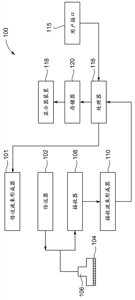
[0024] figure 1 is a schematic diagram of an ultrasound imaging system 100 according to an embodiment. The ultrasound imaging system 100 includes a transmit beamformer 101 and a transmitter 102 that drives elements 104 within a probe 106 to transmit pulsed ultrasound signals into a human body (not shown). The p...


PUM


Abstract

An ultrasound imaging system and method are provided. An ultrasound imaging device (100) includes a probe (106), a display device (118), and a processor (116). The processor is configured to acquire first 4D ultrasound data of the first partial volume (158) for a first period of time that is longer than an estimated cardiac period and to acquire second 4D ultrasound data of the second partial volume (160) for a second period of time that is longer than the estimated cardiac period. The processor is configured to combine the first 4D ultrasound data with the second 4D ultrasound data to generate 4D ultrasound data of a region-of-interest. The processor is configured to display an image generated by the 4D ultrasound data of a region-of-interest.

Description

technical field [0001] The present disclosure generally relates to ultrasound imaging systems and methods for generating 4D ultrasound data of a region of interest by acquiring 4D ultrasound data of multiple local volumes. Background technique [0002] 2D array probes coupled with current generation ultrasound systems are not fast enough to acquire clinically appropriate volume sizes for applications such as fetal heart imaging. Currently, both the required temporal resolution and the required spatial resolution cannot be simultaneously obtained from a single volume acquired with conventional 2D array probes. [0003] Therefore, some conventional ultrasound systems scan slowly across the volume, acquiring 2D image frames at different locations during the scan. For example, for a typical fetal heart rate, the scan may take 8-12 seconds. During scan time, the fetal heart may beat about 16 to 30 times. Phase data indicating the phase of the heart at the time of acquisition o...

Claims


Application Information

Patent Timeline
Patent Type & Authority Applications(China)
IPC IPC(8): A61B8/02A61B5/344
CPCA61B8/488A61B8/486G01S7/52087A61B8/02A61B8/469A61B8/483A61B8/485A61B8/543G01S15/8925A61B8/14A61B8/0866A61B8/0883G01S15/8993G01S7/52065A61B8/466A61B8/145G01S15/89A61B8/00G01S7/52063A61B8/5246A61B8/5253A61B8/5284
Inventor D.J.巴克顿C.F.佩里D.C.赫尼希曼A.肖瓦斯沃尔
Owner GENERAL ELECTRIC CO
Features
  • R&D
  • Intellectual Property
  • Life Sciences
  • Materials
  • Tech Scout
  • Unparalleled Data Quality
  • Higher Quality Content
  • 60% Fewer Hallucinations
Learn More