
A bronchoscopic electrocoagulation probe

A bronchoscopy and electrocoagulation technology, applied in the field of medical devices, can solve problems such as limitations, and achieve the effects of convenient operation, simple structure, and lengthening of the effective electrocoagulation length.

Inactive Publication Date: 2017-11-21
SECOND MILITARY MEDICAL UNIV OF THE PEOPLES LIBERATION ARMY
View PDF2 Cites 0 Cited by
  • Summary
  • Abstract
  • Description
  • Claims
  • Application Information

AI Technical Summary

Problems solved by technology

This invention increases the axial cauterization range of electrocoagulation, but is still limited by the biopsy channel of the bronchoscope in the radial direction

Method used


Image

  • A bronchoscopic electrocoagulation probe
  • A bronchoscopic electrocoagulation probe
  • A bronchoscopic electrocoagulation probe

Examples


Embodiment 1

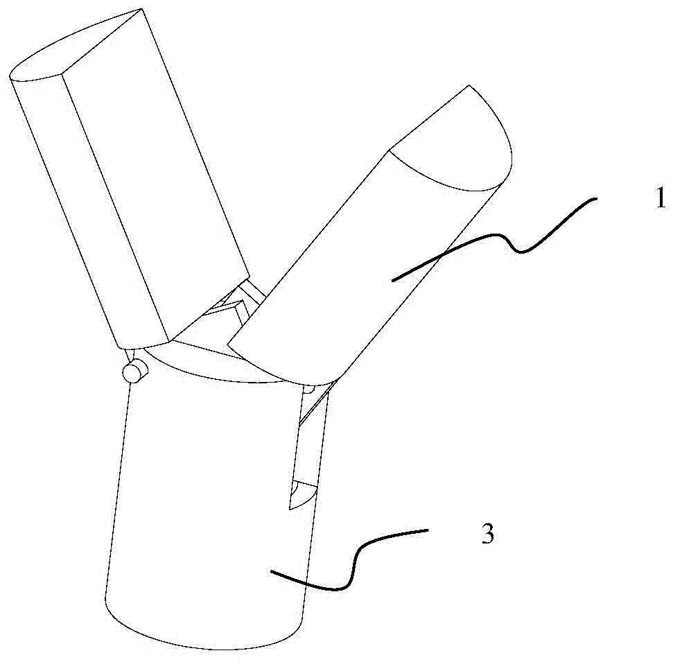
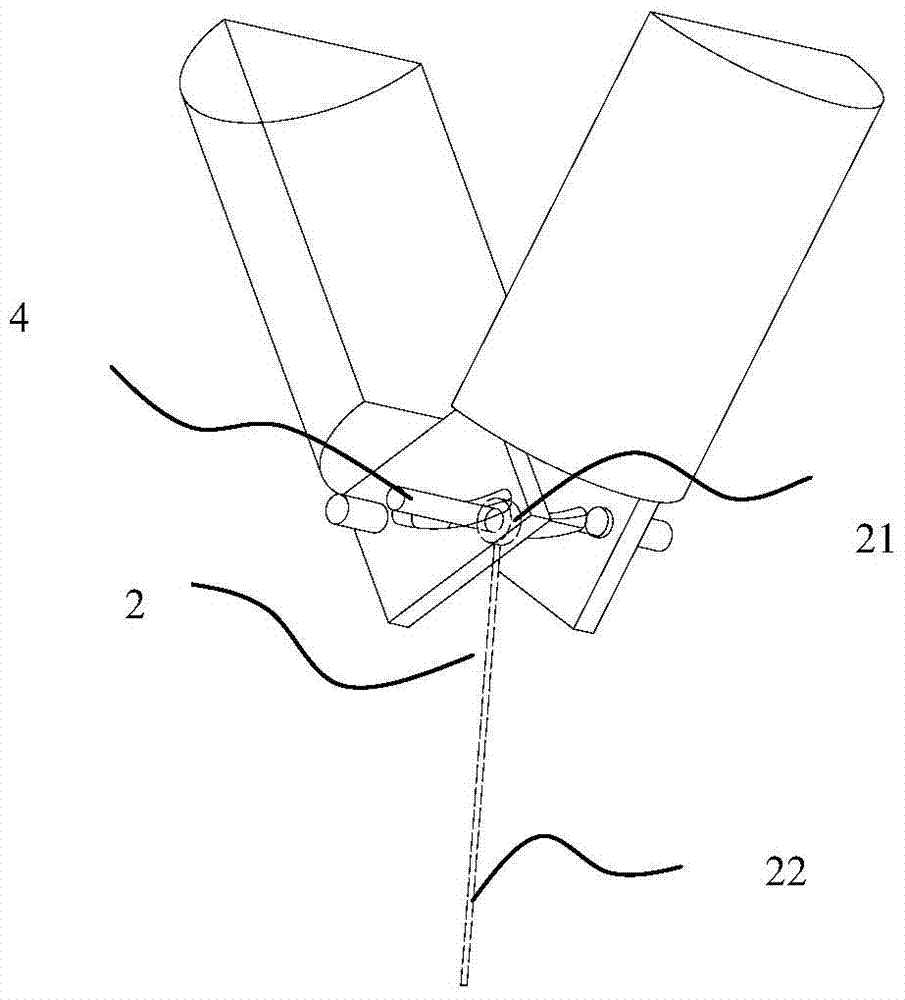
[0026] A bronchoscopic electrocoagulation probe passes through the biopsy channel of the bronchoscope and connects the electrocoagulation wire and the electrodes. The electrocoagulation probe includes two electrodes 1 connected by pivots; the electrodes 1 are also provided with a manipulation Line 2, the control line is composed of a bayonet 21 and a steel wire 22, the bayonet 21 controls the rotation of the two probes, the end of the steel wire 22 is connected to the control handle; the electrode 1 is fixed on the base, the base 3 is hollow, and the electrocoagulation wire and the steel wire are both Through the inner hole of the base 3.

Embodiment 2

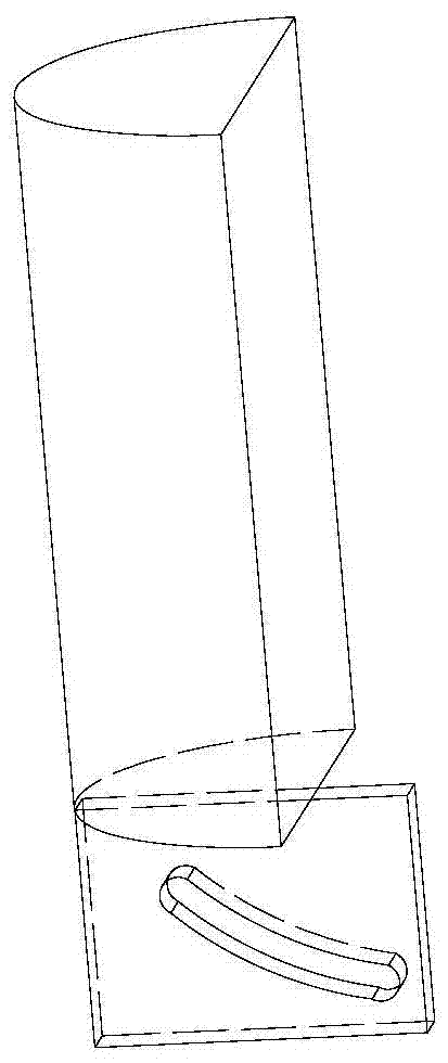
[0028] like Figure 1 ~ Figure 3 As shown, a bronchoscopic electrocoagulation probe has two electrodes 1, both of which are semi-cylindrical. The top of the electrode 1 is used for electrocoagulation, and the bottom is used for connection. The connection part is made into a sheet for easy connection. There are holes on the outside of the electrode connecting part, which are respectively connected to both sides of the base through the connecting piece. Each probe can rotate 0°-180° around the connection point, and the rotation angle of the electrode around the connection point is set at 4 fixed points, each of which is 0° , 60°, 120° and 180°. There is a slideway at the bottom of the electrode connection part, the slideway is curved, and the connecting shaft 4 passes through the slideway 11 of the electrode connection part to form a pivot connection. Since the outer side of the electrode connection part is connected to the base 3, when the connection shaft is pulled to move up...

Embodiment 3

[0030] like Figure 4 A bronchoscopic electrocoagulation probe is shown, and the rest are the same as in embodiment 2, wherein the electrocoagulation probe also includes two connecting pieces 5, the top of the electrode connecting part has holes, and the two electrodes 1 and the base are connected through the connecting piece 3. The electrode rotates around the central support point. The bottom end of the electrode connection part is connected to the connecting piece, and the connecting pieces are connected to each other to form a rhombus, and the bayonet 22 of the manipulation wire 2 is clamped to the connecting piece.


PUM


Abstract

The invention relates to the field of medical equipment, and discloses a bronchoscopic electrocoagulation probe, which passes through the biopsy channel of the bronchoscope and connects the electrocoagulation wire and the electrode, and is characterized in that the electrocoagulation probe includes two probes, two The two probes are pivotally connected; the electrocoagulation probe is also provided with a control line, and the end of the control line is connected to the control handle; the electrocoagulation probe is fixed on the base, the base is hollow, and the electrocoagulation line and the control line pass through the inner hole of the base. The electrocoagulation probe can rotate around the connection point, and the control line moves up and down, and its movement drives the connection point, which can be converted into the rotational movement of the probe to achieve the effect of adjusting the angle of the probe. The present invention changes the cylindrical electrocoagulation probe into two, which can increase the burning range; the two probes are pivotally connected and can rotate around the connection point, and the rotation angle depends on the movement of the control line and the connection relationship between the probe, the base and the control line Adjustment, by pulling the control line, the opening size of the two probes can be adjusted to change the burning range.

Description

technical field [0001] The invention belongs to the field of medical devices, and in particular relates to an electrocoagulation probe used for penetrating a bendable bronchoscope for electrocoagulation during the treatment of benign airway stenosis. Background technique [0002] Benign airway stenosis is a common disease of the respiratory system, which can lead to respiratory failure and even life-threatening in severe cases. It is often necessary to treat the stenosis with a flexible bronchoscope. One of the currently commonly used methods is to electrocoagulate the stenosis through a flexible bronchoscope. The high-frequency current is concentrated on a point in contact with it to generate heat to the local tissue of the human body, and cauterization of the lesion can relieve airway stenosis. The operation requires a bronchoscope, a high-frequency electrocautery instrument and the corresponding electric knife, electrocoagulation probe, snare and other instruments. Durin...

Claims


Application Information

Patent Timeline
Patent Type & Authority Patents(China)
IPC IPC(8): A61B18/12
Inventor 聂小蒙周国武张伟白冲李强王琴王湘奇
Owner SECOND MILITARY MEDICAL UNIV OF THE PEOPLES LIBERATION ARMY
Learn More