Mr imaging using shared information among images with different contrast

A technology of imaging sequence and image representation, which is applied in the direction of using nuclear magnetic resonance imaging system for measurement, magnetic variable measurement, magnetic resonance measurement, etc., to avoid misregistration and improve accuracy.

Inactive Publication Date: 2014-07-16
KONINKLJIJKE PHILIPS NV
View PDF5 Cites 3 Cited by
  • Summary
  • Abstract
  • Description
  • Claims
  • Application Information

AI Technical Summary

Problems solved by technology

Prescan data is processed to create prescan information

Method used


Image

  • Mr imaging using shared information among images with different contrast
  • Mr imaging using shared information among images with different contrast
  • Mr imaging using shared information among images with different contrast

Examples


Embodiment Construction

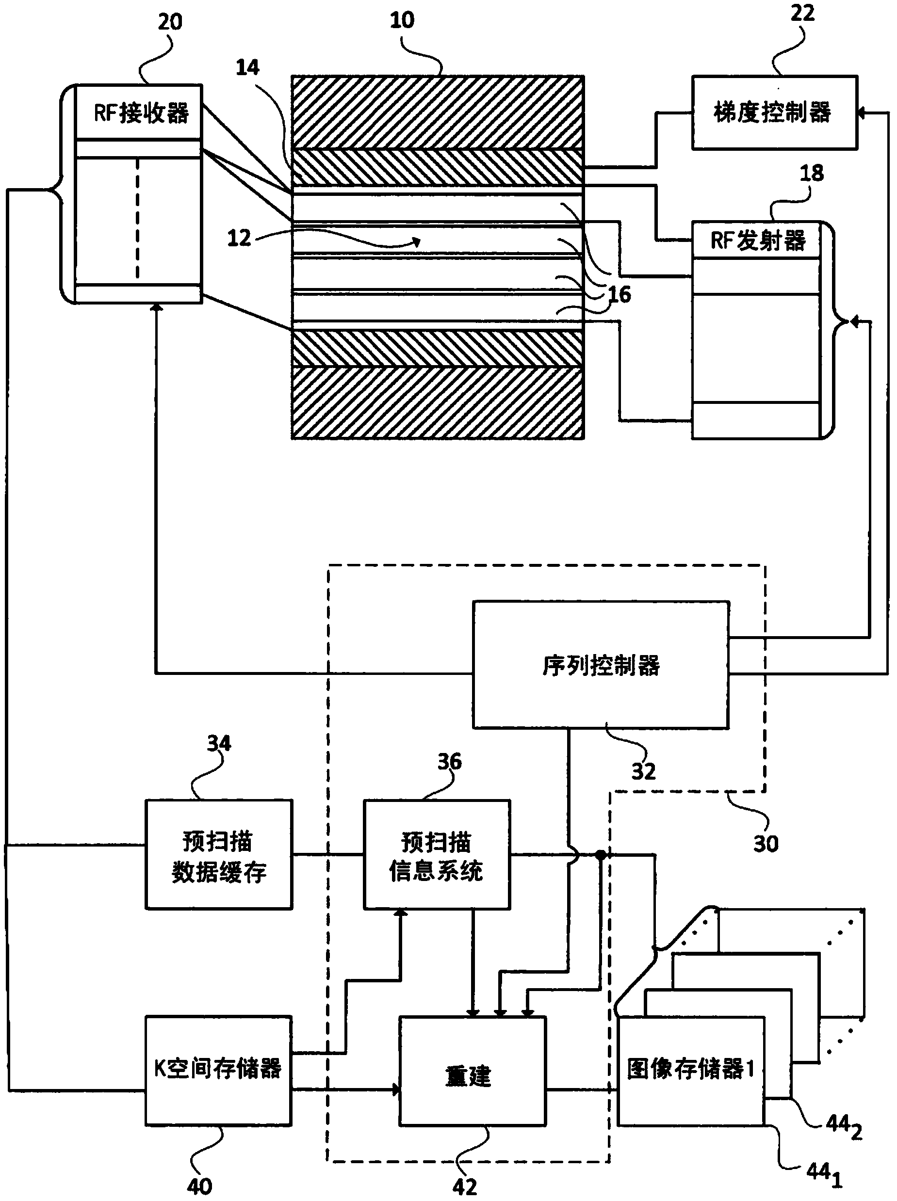
[0026] refer to figure 1 , the magnetic resonance imaging system includes a magnet 10 that generates a static B in the examination region 12 0 field. One or more gradient field magnets 14 generate B across the imaging region 0 The magnetic field gradient of the field. RF coil or element 16 generates B 1 RF pulses are used to stimulate and manipulate magnetic resonance and to induce magnetic resonance signals. Although shown as whole body transmit and receive RF coils, it should be appreciated that separate RF coils may be provided for transmit and receive and that the receive and / or transmit coils may be local coils, whole body coils, or a combination of both. Although shown as a bore type magnetic resonance system, a C-type or open type magnetic resonance system is also contemplated. One or more RF transmitters 18 apply RF signals to the radio frequency coils so that B 1 Pulses are applied in the inspection area. One or more receivers 20 receive magnetism and demodulat...


PUM


Abstract

A typical clinical MR protocol is composed of several sets of scans to acquired images with different contrast, such as T1, T2 and DWI. Currently, the acquisition and reconstruction of these images are processed individually. The proposed method treats the optimization of all acquisitions and reconstructions as one single procedure for faster and more robust MRI. The theory behind this concept is that the information such as B0, B1- field, optimized acquisition trajectory, reconstruction parameters, etc., can be shared among all scans for different contrasts since the same subject is scanned in the same system using the same RF coil.; A method of magnetic resonance imaging includes performing a first magnetic 10 resonance scan sequence which saves a data store, and performing a second magnetic resonance scan sequence which uses a data store from the first magnetic resonance scan sequence. A magnet (10) generates a B0 field in an examination region (12), a gradient coil system (14, 22) creates magnetic gradients in the examination region, and an RF system (16, 18, 20) induces resonance in and receives resonance signals from a subject in the 1 examination region. One or more processors (30) are programmed to perform a magnetic resonance pre-scan sequence to generate pre-scan information, perform a first sequence to generate first sequence data, refine the pre-scan information with the first sequence data, perform a second imaging sequence to generate second sequence data.; Further, the second sequence data is either reconstructed using the refined pre-scan information or performed using the refined pre-scan sequence information.

Description

technical field [0001] This application relates to the field of magnetic resonance (MR). It has particular application in connection with Magnetic Resonance Imaging (MRI) and may also be suitable for Magnetic Resonance Spectroscopy (MRS). Background technique [0002] Magnetic resonance imaging (MRI) uses pre-scans to calibrate and create an initial reference before each scan sequence. A typical pre-scan includes coil survey, sense reference, B0 mapping and B1 mapping. Coil surveys typically last longer than 10 seconds. Sensing references typically last for more than 10 seconds. The B0 mapping lasts more than 15 seconds, and the B1 mapping lasts between 15 and 30 seconds. A total prescan can last longer than one minute. If the coil or patient position changes, the information is inaccurate. In theory, all these pre-scans need to be repeated. Otherwise, the reconstructed image may contain severe artifacts. However, the repetition of these reference scans extends the t...

Claims


Application Information

Patent Timeline
Patent Type & Authority Applications(China)
IPC IPC(8): G01R33/54G01R33/565
CPCG01R33/243G01R33/246G01R33/543G01R33/5611G01R33/565G01R33/56509G01R33/56
Inventor F·黄G·R·丁辛W·林
Owner KONINKLJIJKE PHILIPS NV
Why Eureka
Try Eureka