Method for partitioning breast lesion

A technology of breast and lesion, applied in the field of medical image processing, to achieve the effect of strong stability and robustness

Active Publication Date: 2014-11-12
SHANGHAI UNITED IMAGING HEALTHCARE
View PDF4 Cites 16 Cited by
  • Summary
  • Abstract
  • Description
  • Claims
  • Application Information

AI Technical Summary

Problems solved by technology

[0005] The problem to be solved by the present invention is to provide a method for segmenting breast lesions, which is used to solve the problem in

Method used


Image

  • Method for partitioning breast lesion
  • Method for partitioning breast lesion
  • Method for partitioning breast lesion

Examples


Embodiment Construction

[0038] In the following description, numerous specific details are set forth in order to provide a thorough understanding of the present invention. However, the present invention can be implemented in many other ways different from those described here, and those skilled in the art can make similar extensions without violating the connotation of the present invention, so the present invention is not limited by the specific implementations disclosed below.

[0039] Secondly, the present invention is described in detail by means of schematic diagrams. When describing the embodiments of the present invention in detail, for convenience of explanation, the schematic diagrams are only examples, which should not limit the protection scope of the present invention.

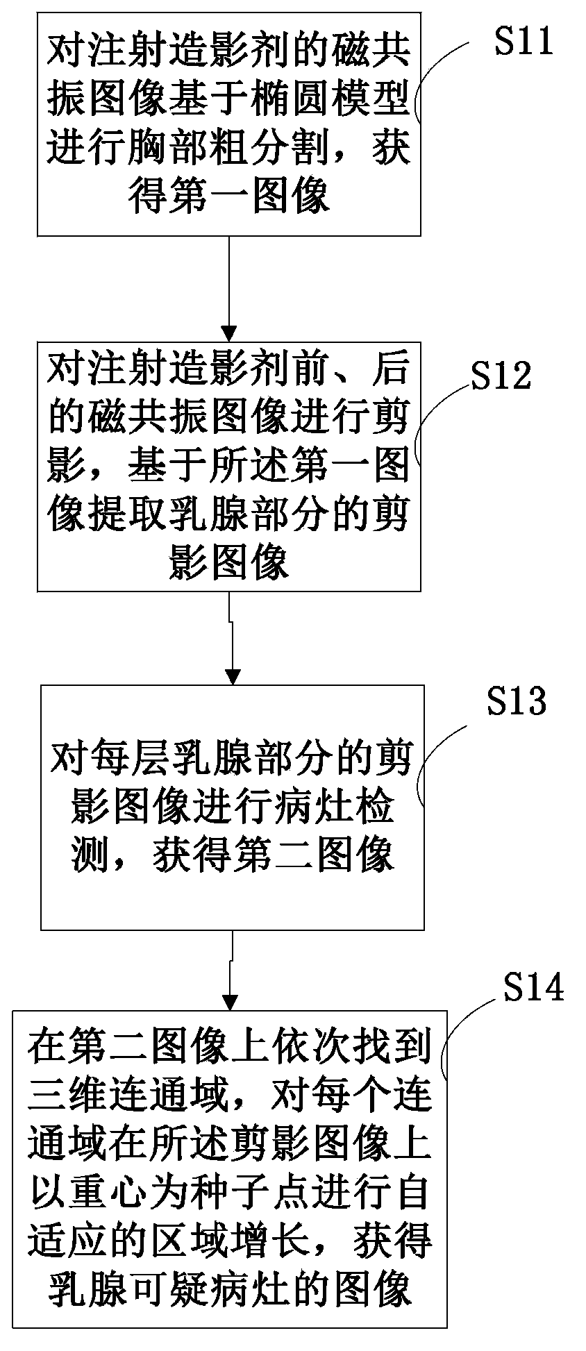
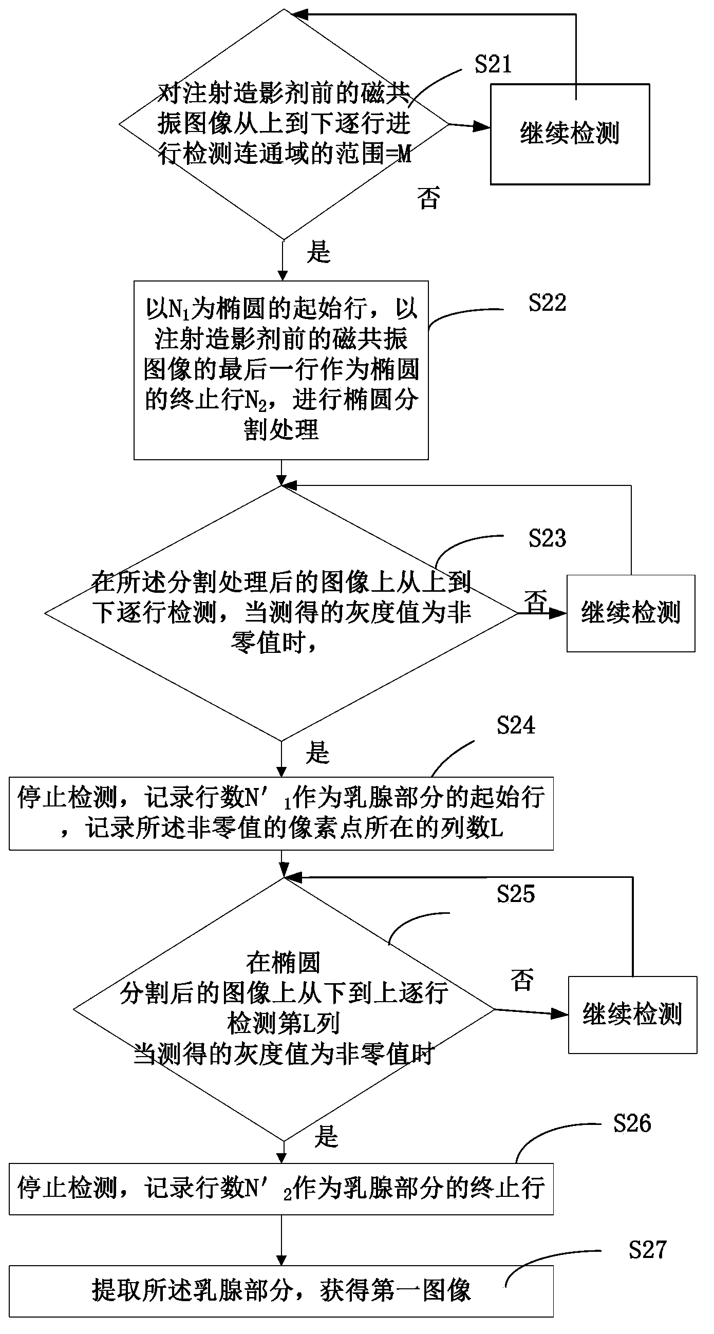
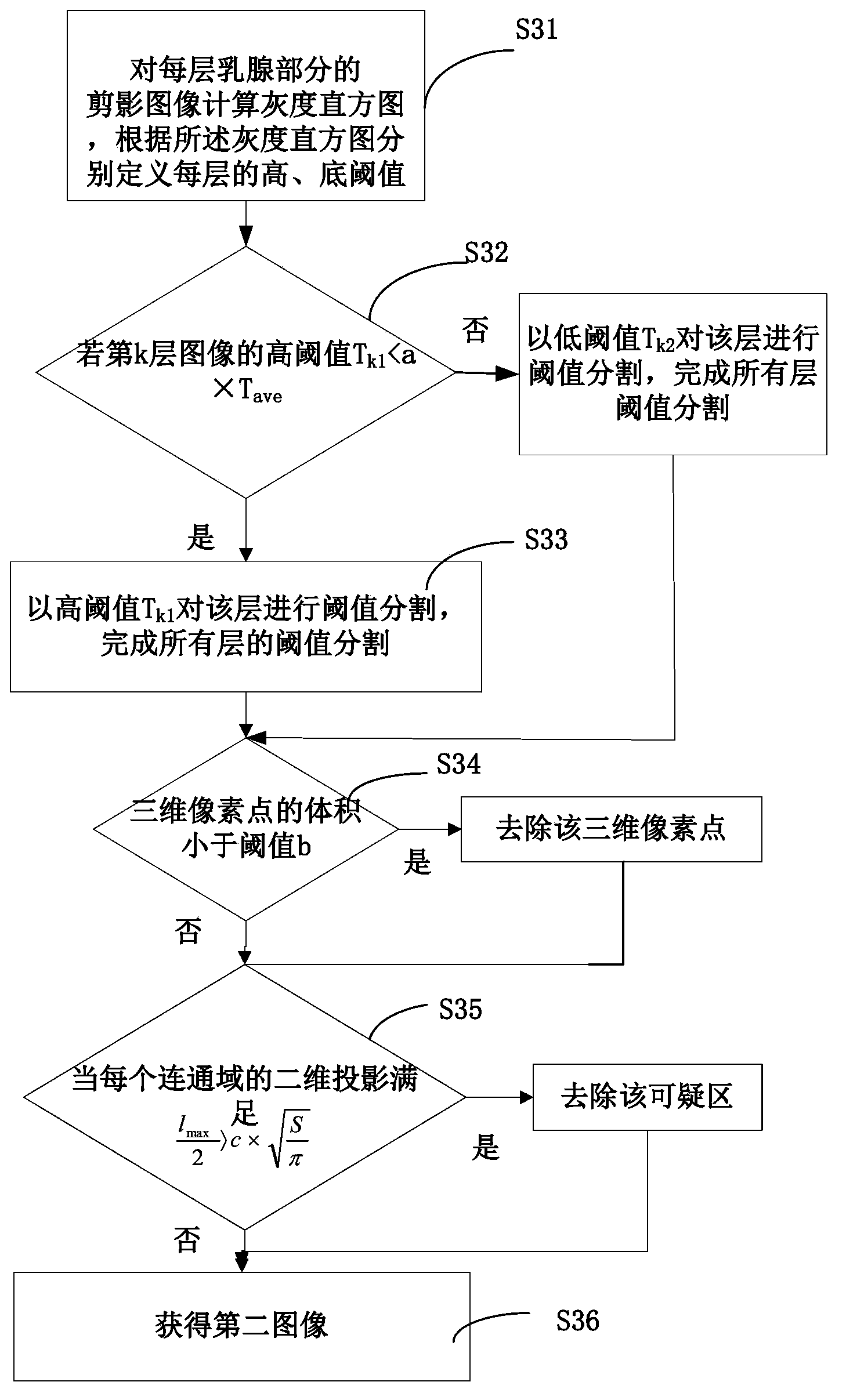
[0040] In order to solve the technical problems in the background art, this embodiment provides a method for segmenting breast lesions. figure 1 Shown is a schematic flow chart of a method for segmenting breast lesions in...


PUM


Abstract

The invention provides a method for partitioning breast lesion. The method comprises steps as follows: coarse chest partition is performed on a magnetic resonance image on the basis of an oval model before a contrast medium is injected, and a first image is acquired; silhouette is performed after registration of magnetic resonance images obtained before and after the contrast medium is injected, and a silhouette image of the breast part is extracted from the acquired silhouette images on the basis of the first image; the silhouette image of each layer of breast parts is subjected to lesion detection, and a second image is acquired; and a three-dimensional communication area is found sequentially from the second image, self-adaptive region growing is performed on the silhouette image by taking the gravity center of each communication area as a seed point, and the magnetic resonance images are acquired after suspicious lesions of the breast are partitioned. The provided method for partitioning breast lesions can simply, effectively and automatically partition the suspicious lesions and can be widely applied to magnetic resonance imaging data by injecting various contrast media.

Description

technical field [0001] The invention relates to the field of medical image processing, in particular to a method for segmenting breast lesions. Background technique [0002] Computer Aided Diagnosis (CAD) refers to the use of imaging, medical image processing technology and other possible physiological and biochemical means, combined with computer analysis and calculation, to assist radiologists in finding lesions and improving the accuracy of diagnosis. With the development of technology, modern high-definition imaging equipment represented by magnetic resonance imaging and computerized tomography provides great convenience for the diagnosis of clinical diseases. At present, CAD research is relatively mature in breast and lung lesions; [0003] Breast cancer is one of the most common cancers in women and the second leading cancer killer in women. Magnetic resonance imaging has become an effective means of assisting doctors in detecting breast cancer because of its high se...

Claims


Application Information

Patent Timeline
IPC IPC(8): G06F19/00G06T7/00
Inventor 李华
Owner SHANGHAI UNITED IMAGING HEALTHCARE
Why Eureka
Try Eureka