Method of, and apparatus for, processing medical image

A processing device and medical image technology, applied in image data processing, image enhancement, image analysis, etc., can solve problems such as complex methods, time-consuming and labor-intensive, difficult to select angles and measurement angles of view

Active Publication Date: 2014-12-24
TOSHIBA MEDICAL SYST CORP
View PDF3 Cites 6 Cited by
  • Summary
  • Abstract
  • Description
  • Claims
  • Application Information

AI Technical Summary

Problems solved by technology

However, this can take time and effort
The visibility of the lumen and the evaluation ability of the narrow portion vary depending on the angle, so it is difficult to select the angle and measure the angle of view
In addition, identification and quantification based on manual work may also p

Method used


Image

  • Method of, and apparatus for, processing medical image
  • Method of, and apparatus for, processing medical image
  • Method of, and apparatus for, processing medical image

Examples


Embodiment Construction

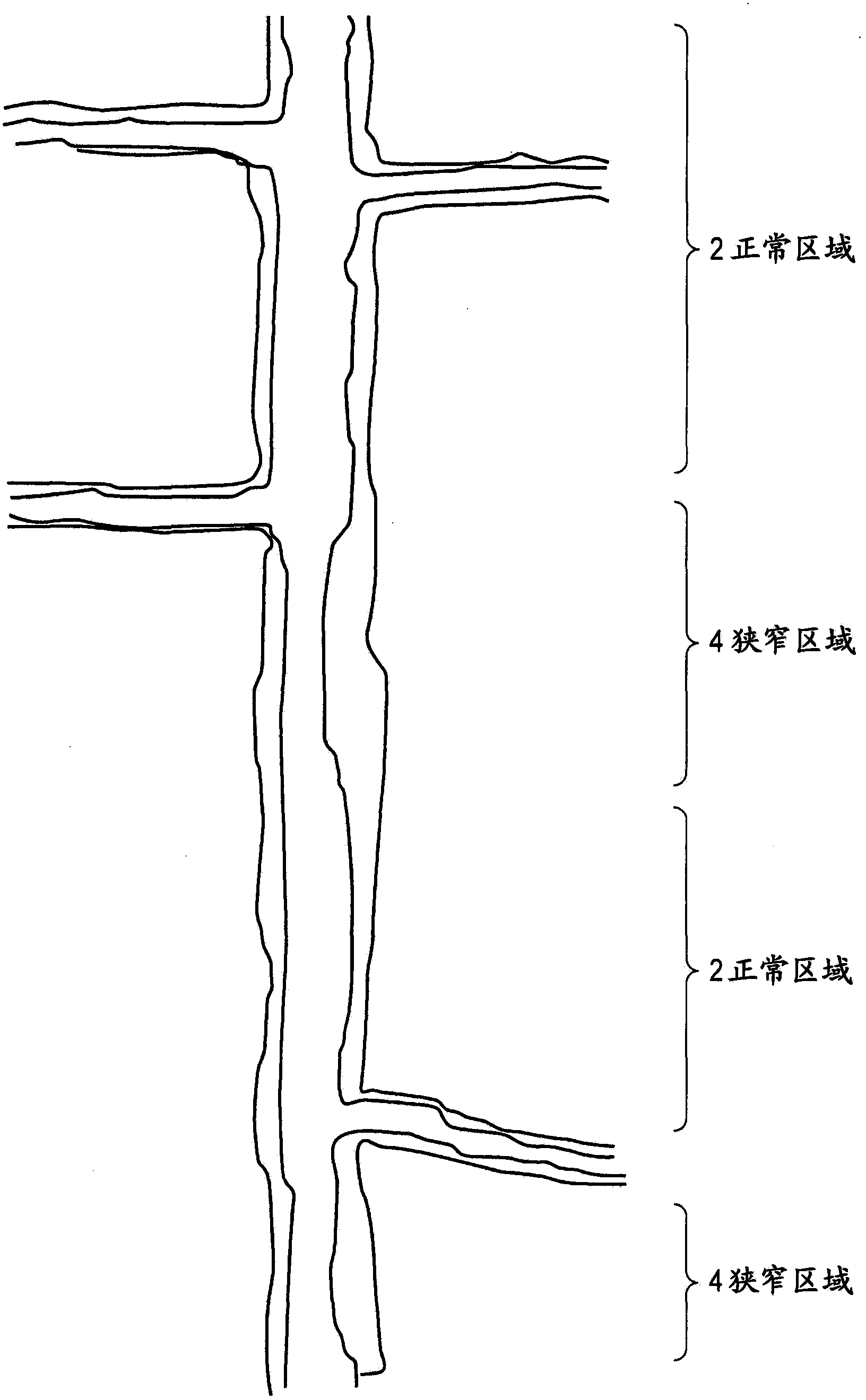
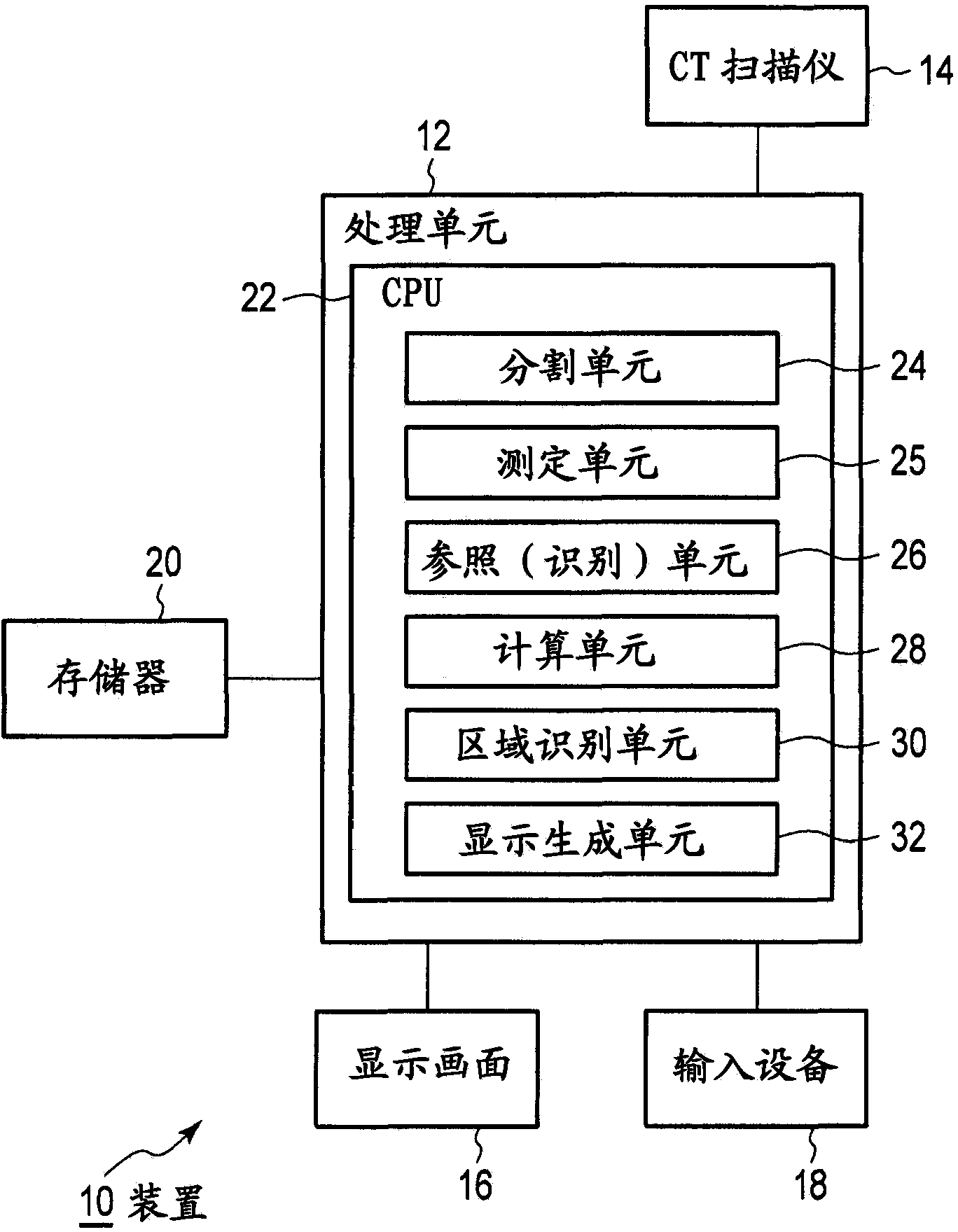
[0019] Several embodiments provide an apparatus for processing volume data to identify a lumen region having a prescribed state, the apparatus comprising: a measurement unit for measuring at least one lumen parameter; lumen data, identifying at least 1 reference segment in the non-branched lumen; the calculation unit, based on the measured value of at least 1 parameter in the at least 1 reference segment, calculates the expected value of the parameter in a further segment of the lumen; The area identifying unit identifies at least one lumen area having a predetermined state dependent on both the expected value of the parameter in the further segment and the measured value of the parameter in the further segment.

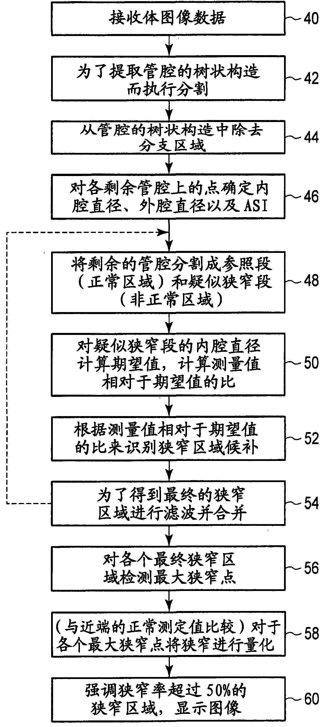
[0020] Several embodiments provide a method of automatically detecting a lumen region, the method comprising: obtaining data representing a non-branching lumen; identifying at least one reference segment of the non-branching lumen; and measuring at least one reference...


PUM


Abstract

The invention provides a medical image processing apparatus capable of assessing each lumen in unbranched vessel sections of the lumen, and automatically identifying whether a narrow palace exists. The related medical image processing apparatus comprises a segmentation unit, a measurement unit, a reference identification unit, a determination unit, an expected value calculation unit and an identification unit. The segmentation unit is used for dividing a vessel tree in to a plurality of unbranched vessel sections through branch region of vessel tree in the lumen in volumetric data. The measurement unit is used for measuring characteristic parameters relative to a cross-section shape of the lumen in each unbranched vessel section. The index calculation unit is used for calculating lumen state indexes representing each unbranched vessel section according to the characteristic parameters. The determination unit is used for determining an abnormal region and a normal region in each unbranched vessel section according to the lumen state indexes. The expected value calculation unit is used for calculating an expected value of a characteristic parameter of the abnormal region according to a characteristic parameter of the normal region adjacent to the abnormal region. The identification unit is used for identifying whether a narrow palace exists in the abnormal region according to a measured value of the characteristic parameter of the abnormal region and the expected value.

Description

technical field [0001] Embodiments described herein generally relate to methods and apparatus for processing stereoscopic image data to identify lumen regions having a defined state. Embodiments have utility in the identification and quantification of stenosis within a lumen. Background technique [0002] In order to obtain image data representing the internal structure of a patient or other subject, it is known to use medical image diagnosis technology. There is known an angiography method in which blood vessels are imaged. [0003] Conventionally, angiography has been in the form of catheter angiography, in which a catheter is inserted into a large lumen. Next, the catheter is induced to the point of interest (eg, the heart). At this point, a contrast agent is injected from the point of interest. Afterwards, multiple two-dimensional still images, or short animated images, are taken using X-ray fluoroscopy. In such images, the lumen can be clearly seen through the cont...

Claims


Application Information

Patent Timeline
IPC IPC(8): A61B6/03
CPCG06T7/0012G06T2207/10081G06T2207/10088G06T2207/30101G06T2207/30172A61B5/026A61B5/02007A61B5/7275
Inventor C·普拉卡斯B·莫赫S·马苏德
Owner TOSHIBA MEDICAL SYST CORP
Why Eureka
Try Eureka