Intravascular ultrasound image and intravascular-OCT image fusing method

A fusion method, IV-OCT technology, applied in image enhancement, image data processing, instruments, etc.

Active Publication Date: 2015-02-25
NORTH CHINA ELECTRIC POWER UNIV (BAODING)
View PDF3 Cites 29 Cited by
  • Summary
  • Abstract
  • Description
  • Claims
  • Application Information

AI Technical Summary

Problems solved by technology

However, so far, people have not been able to f...

Method used


Image

  • Intravascular ultrasound image and intravascular-OCT image fusing method
  • Intravascular ultrasound image and intravascular-OCT image fusing method
  • Intravascular ultrasound image and intravascular-OCT image fusing method

Examples


Embodiment Construction

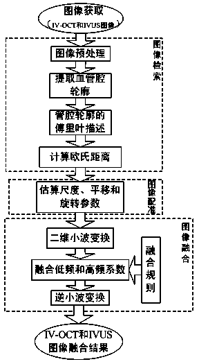
[0055] The present invention provides a method for automatic fusion of intravascular ultrasound gray-scale images and intravascular OCT images. The data processing steps of the present invention will be described in detail below in conjunction with the accompanying drawings:

[0056] 1. Image retrieval

[0057] When performing IVUS and IV-OCT imaging on the same vessel, the frame rate of IV-OCT is frame / s, the catheter retraction speed is mm / s; the frame rate of IVUS is frame / s, the catheter retraction speed is mm / s. Therefore, the number of IVUS image frames collected in the retraction distance of 1mm is , the number of IV-OCT image frames is , the ratio between the two is . For example, the frame rate of IV-OCT is 100 frames / s, and the catheter retraction speed is 20 mm / s; the frame rate of IVUS is 30 frames / s, and the catheter retraction speed is 0.5 mm / s. Then the number of IVUS image frames collected in a retraction distance of 1mm is 12 times that of IV-OC...


PUM


Abstract

The invention relates to an intravascular ultrasound image and intravascular-OCT image fusing method. The method includes the following steps of (a) image retrieval, wherein for intravascular ultrasound (IVUS) images and intravascular (IV)-OCT images collected at the same position on a blood vessel section, a frame of IV-OCT image is taken as a reference image, and an image to be registered is selected from the n frames of IVUS images collected from the point; (b) IVUS image and IV-OCT image registration; (c) IVUS image and IV-OCT image fusion. IV-OCT image data and IVUS image data of the same blood vessel section are fused, the advantage of high tissue penetrating power of IVUS imaging and the advantage of high resolution of IV-OCT imaging are brought into full play, more comprehensive description of blood vessel walls and atherosclerotic plaques is obtained, and a reliable basis is provided for research on the coronary heart disease and the like.

Description

technical field [0001] The present invention relates to a method for automatically fusing intravascular ultrasound (intravascular ultrasound, IVUS) gray-scale images and intravascular OCT (intravascular optical coherence tomography, IV-OCT) images of the same segment of blood vessels, so as to fully display the vascular lumen and blood vessels The invention relates to the morphological structure of wall (including atherosclerotic plaque) tissue, belonging to the technical field of medical imaging. Background technique [0002] Intravascular ultrasound (IVUS) is an interventional imaging method commonly used in clinical research on vascular lesions. It places a miniature high-frequency ultrasound probe on the top of the cardiac catheter, and uses the cardiac catheter to guide the ultrasound probe into the vascular cavity for detection, and then the electronic imaging system To display the micro-anatomical information of the tissue structure and geometry of the vessel wall. I...

Claims


Application Information

Patent Timeline
IPC IPC(8): G06T5/50
Inventor 孙正胡宏伟
Owner NORTH CHINA ELECTRIC POWER UNIV (BAODING)
Why Eureka
Try Eureka