Liver boundary identification method and system

A boundary recognition and boundary technology, applied in the liver boundary recognition method and system field, can solve the problems of high operator requirements, long recognition time, low efficiency of liver boundary recognition, etc., and achieve the effect of improving recognition efficiency and efficient recognition

Active Publication Date: 2015-03-11
WUXI HISKY MEDICAL TECH
View PDF4 Cites 9 Cited by
  • Summary
  • Abstract
  • Description
  • Claims
  • Application Information

AI Technical Summary

Problems solved by technology

Due to the method of manually selecting the liver boundary based on the information of the liver tissue, the operator needs to be very familiar with the structure of the liver tissue and image information in order to accurately select the boundary of the liver tissue, so the operator has high requirements; at the same time, due to the Through manual identification, the identification time is longer, resulting in lower identification efficiency of the liver boundary

Method used


Image

  • Liver boundary identification method and system
  • Liver boundary identification method and system
  • Liver boundary identification method and system

Examples


no. 1 example

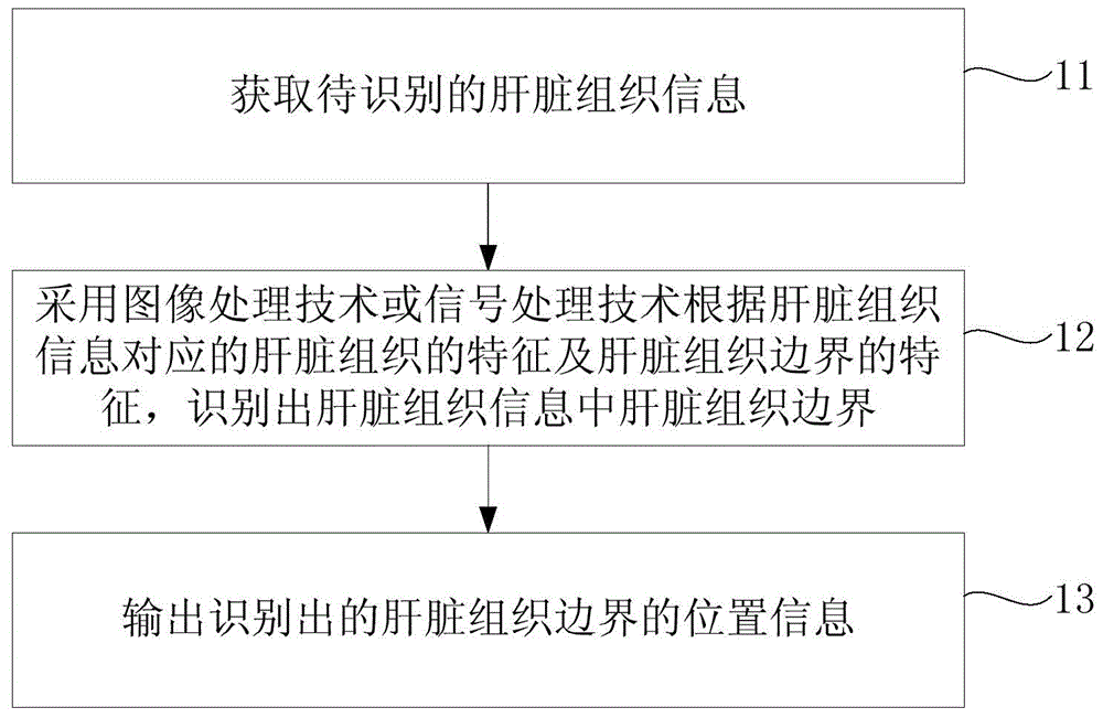
[0024] figure 1 It is an implementation flowchart of the liver boundary identification method provided in the first embodiment of the present invention, and the method can be executed by the liver boundary identification system. Such as figure 1 As shown, the implementation process includes:

[0025] Step 11. Obtain the liver tissue information to be identified.

[0026] Wherein, the liver tissue information may be an A-mode ultrasonic signal of the liver tissue, an M-mode ultrasonic signal of the liver tissue, a B-mode ultrasonic image of the liver tissue, a CT image of the liver tissue or an MRI image of the liver tissue.

[0027] According to the liver tissue information, the type of liver tissue information can be obtained, that is, the type of liver tissue information can be ultrasonic signals, such as A-mode ultrasonic signals of liver tissue and M-mode ultrasonic signals of liver tissue, or two-dimensional ultrasonic images, such as liver The B-mode ultrasound image ...

no. 2 example

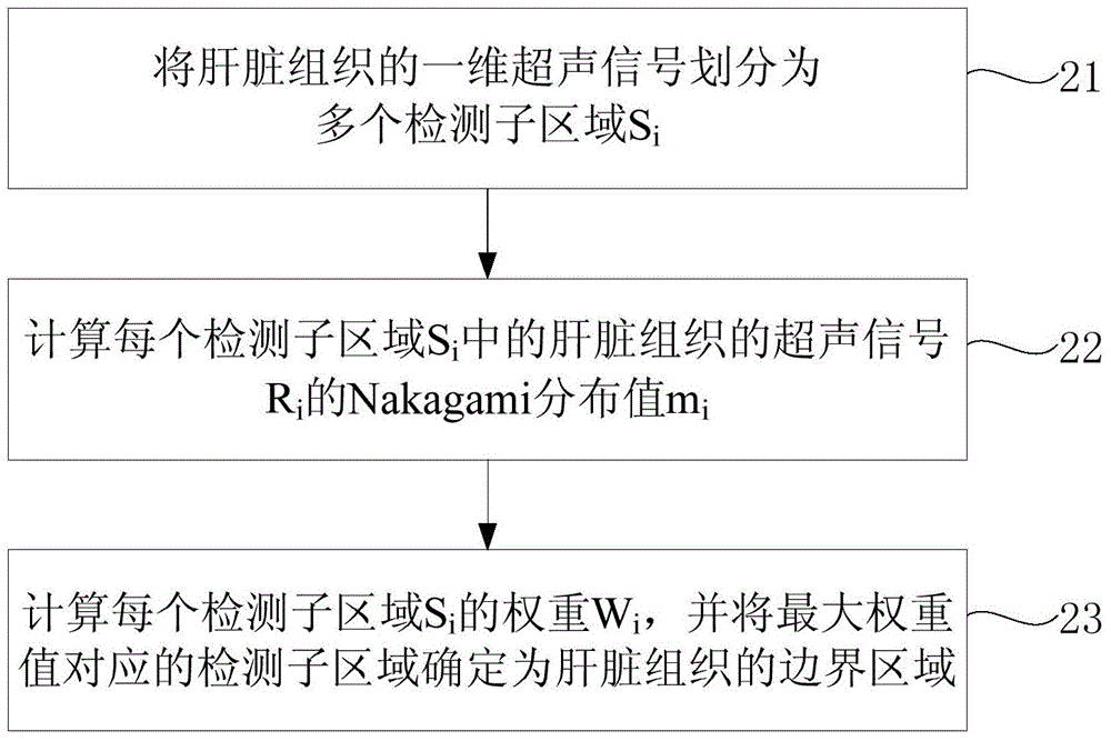
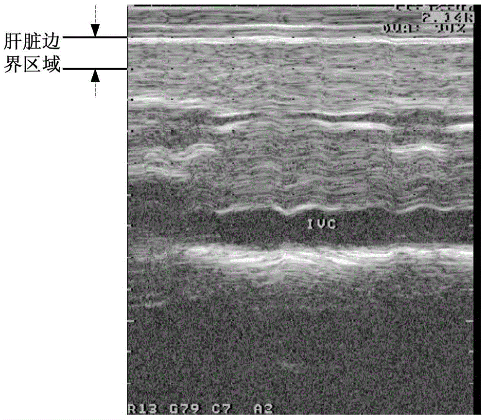
[0035] The second embodiment is a specific optimization of step 12 in the first embodiment of the present invention, and this method is applicable to one-dimensional ultrasound signals of liver tissue. figure 2 is an implementation flowchart of the liver boundary recognition method provided in the second embodiment of the present invention, image 3 It is an effect diagram of boundary recognition based on M-mode ultrasound signals of liver tissue in the second embodiment of the present invention. combine figure 2 and image 3 , the method includes:

[0036] Step 21. Divide the one-dimensional ultrasound signal of the liver tissue into multiple detection sub-regions S i .

[0037] The one-dimensional ultrasound signal of the liver tissue may be an A-mode ultrasound signal of the liver tissue or an M-mode ultrasound signal of the liver tissue. Assuming that an ultrasound signal contains n sampling points, and the scanning depth of the corresponding ultrasound signal of th...

no. 3 example

[0051] The third embodiment is a specific optimization of step 12 in the first embodiment of the present invention, and this method is applicable to two-dimensional ultrasound images of liver tissue. Figure 4 is an implementation flowchart of the liver boundary recognition method provided in the third embodiment of the present invention, Figure 5 It is an effect diagram of boundary recognition based on a B-mode ultrasound image of liver tissue in the third embodiment of the present invention. combine Figure 4 and Figure 5 , the method includes:

[0052] Step 31. Divide the two-dimensional ultrasound image of the liver tissue into a plurality of rectangular detection sub-regions R ij .

[0053] The two-dimensional ultrasound image of the liver tissue may be a B-mode ultrasound image of the liver tissue. Assuming that the size of a B-ultrasound image is w*h, where w is the width of the two-dimensional ultrasound image of the liver tissue, h is the height of the two-dime...


PUM


Abstract

The embodiment of the invention relates to the technical field of medical image processing, in particular to a liver boundary identification method and system. The method comprises the following steps: obtaining liver tissue information to be identified; adopting an image processing technology or a signal processing technology to identify a liver tissue boundary in the liver tissue information according to the characteristics of a liver tissue corresponding to the liver tissue information and the characteristics of the liver tissue boundary; and outputting the position information of the identified liver tissue boundary. When the method is adopted, the liver tissue boundary can be automatically identified, and liver boundary identification efficiency can be improved so as to realize the automatic positioning of the liver boundary.

Description

technical field [0001] The embodiments of the present invention relate to the technical field of medical image processing, and in particular to a method and system for identifying liver boundaries. Background technique [0002] Many clinical applications need to identify liver tissue boundaries from traditional medical imaging, including ultrasound imaging, magnetic resonance imaging (MRI, Magnetic Resonance Imaging), computed tomography (CT, Computed Tomography), etc. Positioning, such as liver elasticity detection and liver color Doppler ultrasonography, etc. [0003] At present, the boundaries of liver tissue are mainly identified artificially. Due to the method of manually selecting the liver boundary based on the information of the liver tissue, the operator needs to be very familiar with the structure of the liver tissue and image information in order to accurately select the boundary of the liver tissue, so the operator has high requirements; at the same time, due to...

Claims


Application Information

Patent Timeline
Patent Type & Authority Applications(China)
IPC IPC(8): G06K9/00
CPCG06T7/13G06T2207/30056G06T2207/10132G06T7/136A61B8/0858A61B8/5223G06T7/0012G06T2207/20076G06T2207/30008G06T2207/30012G06T7/12G06T7/187G06T7/11G06T2207/10081G06T2207/10088G16H50/30G06F2218/08G06T7/10G16H30/20G06F18/00
Inventor 邵金华段后利孙锦
Owner WUXI HISKY MEDICAL TECH
Why Eureka
Try Eureka