
Medical image data processing apparatus and method

A medical image and processing device technology, applied in image data processing, image data processing, image analysis, etc., can solve the problems of longer processing time and increased calculation load, and achieve the effect of shortening the processing time

Active Publication Date: 2015-05-20
TOSHIBA MEDICAL SYST CORP
View PDF7 Cites 5 Cited by
  • Summary
  • Abstract
  • Description
  • Claims
  • Application Information

AI Technical Summary

Problems solved by technology

However, in this case, the calculation load increases, and there is a possibility that the processing time may become longer.

Method used


Image

  • Medical image data processing apparatus and method
  • Medical image data processing apparatus and method
  • Medical image data processing apparatus and method

Examples


Embodiment Construction

[0027] Hereinafter, a medical image processing device and a medical image processing method according to the present embodiment will be described with reference to the drawings.

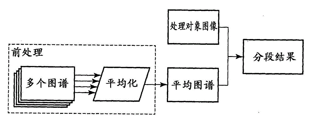
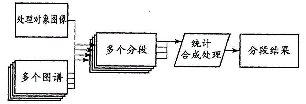
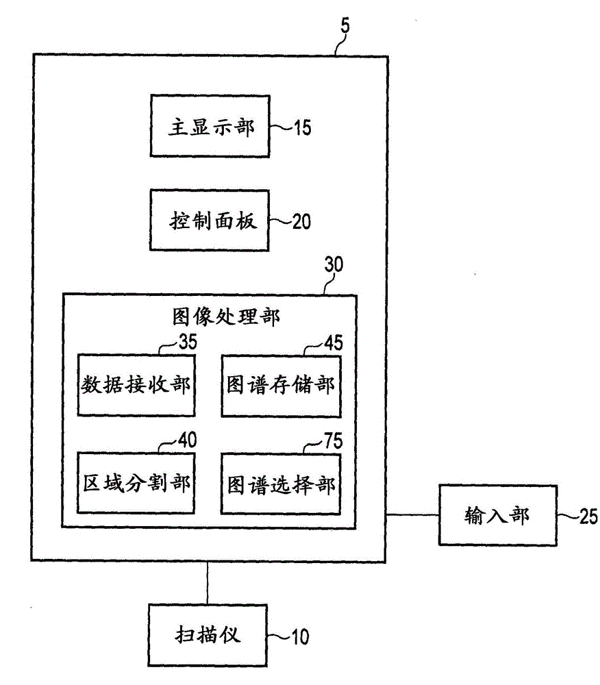
[0028] The medical image processing device according to this embodiment includes: a storage unit that stores a plurality of atlases related to the anatomical atlas of the target site; The positions of the points are identified, and a plurality of selection maps used for segmenting the image to be processed are selected from the plurality of maps.

[0029] The atlas selection unit performs first registration between each atlas of the plurality of atlases and the image to be processed. In addition, the atlas selection unit performs a second registration between the plurality of selected atlases and the image to be processed.

[0030] The calculation amount of the first registration is less than that of the second registration. The first registration is, for example, linear registration. The second r...


PUM


Abstract

An image data processing apparatus comprises a data receiving unit for receiving image data to be segmented, and an atlas selection unit for accessing a plurality of atlas data sets and selecting a subset of the atlas data sets for use in segmenting the image data, wherein the atlas selection unit is configured to select the subset of atlas data sets in dependence on the positions of one or more anatomical landmarks comprised in the plurality of atlas data sets.

Description

[0001] This application claims priority to U.S. Patent Application No. 14 / 079,908 filed on November 14, 2013, and the entire contents of the above U.S. Patent Application are cited in this application. technical field [0002] Embodiments of the present invention relate to a medical image processing device and a medical image processing method. Background technique [0003] A recent medical imaging method generates and analyzes a plurality of two-dimensional, three-dimensional, and four-dimensional images from data collected by a medical image diagnostic apparatus. Such images can be generated using various medical imaging methods such as CT, MRI, PET, and ultrasonic imaging, or medical modality. [0004] Software is used to generate three-dimensional medical images in order to combine measurement data taken at various positions or angles, and to render images from the combined data using methods such as simple surface shading or direct volume rendering. In a four-dimension...

Claims


Application Information

Patent Timeline
IPC IPC(8): G06T1/00G06T7/00
CPCG06T7/11G06T7/174G06T2207/10081G06T2207/10088G06T2207/10104G06T2207/10136G06T2207/20128G06T2207/30008G06T2207/30016G06T2207/30101
Inventor B·莫赫I·普尔
Owner TOSHIBA MEDICAL SYST CORP
Learn More