Computer-assisted lump detecting method based on mammary gland magnetic resonance image

A magnetic resonance image, computer-aided technology, applied in the field of medical image processing and pattern recognition, can solve the problems of poor tumor segmentation, low accuracy, low sensitivity and specificity

Active Publication Date: 2015-06-24
SUN YAT SEN UNIV
View PDF4 Cites 26 Cited by
  • Summary
  • Abstract
  • Description
  • Claims
  • Application Information

AI Technical Summary

Problems solved by technology

[0006] In order to solve the problems of poor segmentation of tumors in the prior art and low accuracy, sensitivity and specificity in classification experiments, the present invention provides a computer-aided tumor detection method based on breast magnetic resonance images. It has a g

Method used


Image

  • Computer-assisted lump detecting method based on mammary gland magnetic resonance image
  • Computer-assisted lump detecting method based on mammary gland magnetic resonance image
  • Computer-assisted lump detecting method based on mammary gland magnetic resonance image

Examples


Embodiment 1

[0106] A computer-aided mass detection method based on breast magnetic resonance images, comprising the following steps:

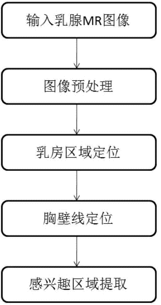
[0107] Such as figure 1 As shown, S1, extracting the region of interest from the breast magnetic resonance image; the specific steps are:

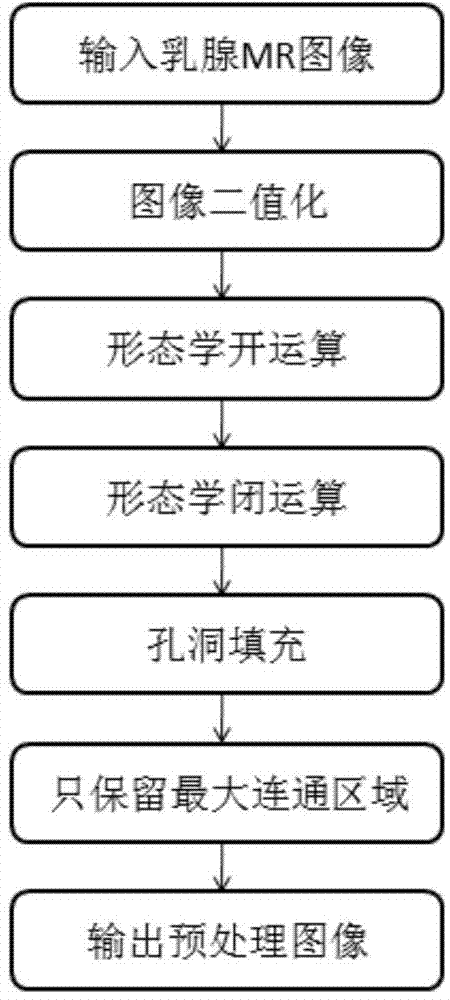
[0108] Such as figure 2 As shown, S11, preprocessing the breast magnetic resonance image to obtain a preprocessing image; it specifically includes:

[0109] S111, perform image binarization processing,

[0110] S112. Perform a morphological opening operation;

[0111] S113. Perform a morphological closing operation;

[0112] S114. Carry out hole filling;

[0113] S115. Extract the output of the largest connected region to obtain the preprocessed image.

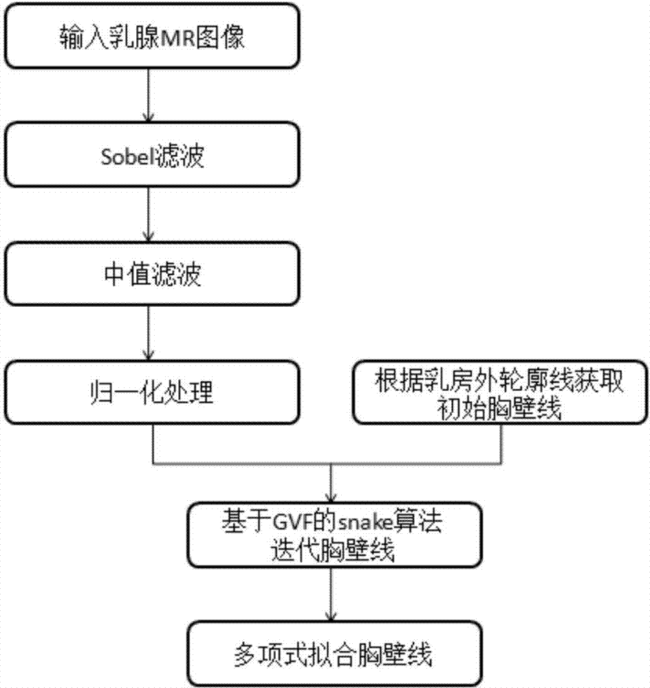
[0114] S12. Extracting the outer contour of the breast; it specifically includes:

[0115] S121. Using an edge operator to extract the outer contour of the breast;

[0116] S122. Perform polynomial fitting on the outer contour of the breast.

[0117] Such as ...

Embodiment 2

[0207] Such as Figure 5 As shown, this embodiment is an improvement made on the basis of the first embodiment, and its difference from the first embodiment is that the outline of the initial tumor area is extracted by using the fuzzy C-means clustering method. It specifically includes:

[0208] S21. Input the region of interest;

[0209] S22. Perform a neighborhood suppression operation;

[0210] S23. Perform a Gaussian denoising filtering operation;

[0211] S24. Perform a histogram equalization operation;

[0212] S25. Perform fuzzy C-means clustering operation;

[0213] S26. Obtain a binarized image;

[0214] S27. Perform hole filling operation;

[0215] S28. Remove the small area and output, that is, obtain, the contour line of the initial mass area.

Embodiment 3

[0217] This embodiment is an improvement made on the basis of Embodiment 2. The difference between it and Embodiment 2 is that: using the result of obtaining the outline of the initial tumor area using the fuzzy C-means clustering method in Embodiment 2 as a basis, using the method based on The snake energy model segmentation method of the gradient vector field performs secondary segmentation and extraction on the contour line of the initial tumor region to obtain the optimized contour line of the initial tumor region.


PUM


Abstract

The invention relates to the field of medical image processing and pattern recognition, and provides a computer-assisted lump detecting method based on a mammary gland magnetic resonance image. The computer-assisted lump detecting method based on the mammary gland magnetic resonance image aims at solving the problems that in the prior art, the lump partition effect is not good, and the accuracy, the sensitivity and the specificity in a classification test are not high. The computer-assisted lump detecting method includes the following steps: S1, extracting an interest area from the mammary gland magnetic resonance image; S2, extracting an initial lump area from the interest area in a separated mode, and determining the contour line of the initial lump area; S3, calculating the weight distribution of characteristic parameters of the initial lump area; S4, selecting the characteristic parameters, with the weight coefficients larger than a standard weight coefficient, of the initial lump area, and carrying out training classifying to obtain optimized characteristic parameters; S5, inputting the optimized characteristic parameters into a classifier, analyzing the optimized characteristic parameters with a support vector machine classification method, determining a final lump area, and displaying the final lump area for a user. The detecting method has the good lump partition effect, the accuracy, the sensitivity and the specificity in the classification test are effectively improved, the detecting result serves as a second opinion to be provided for a radiologist, and the misdiagnosis rate and the missed diagnosis rate of the radiologist can be effectively reduced.

Description

technical field [0001] The invention relates to the fields of medical image processing and pattern recognition, in particular to a computer-aided mass detection method based on breast magnetic resonance images. Background technique [0002] Breast cancer is a malignant tumor that seriously affects women's physical and mental health. According to statistics, its incidence rate accounts for 7-10% of all kinds of malignant tumors in women's whole body. The etiology of breast cancer is not yet fully understood, and there is no good means of prevention and treatment. However, clinical experience shows that the cure rate of early-stage breast cancer patients is much higher than that of middle-advanced patients. Therefore, accurate early diagnosis is the key to reducing the incidence and mortality of breast cancer. At present, the important basis for the diagnosis of breast tumors in the medical field is breast magnetic resonance images, but breast magnetic resonance images are re...

Claims


Application Information

Patent Timeline
IPC IPC(8): G06K9/00G06K9/54G06K9/62G06T7/00
Inventor 庞志勇陈弟虎朱冬梅
Owner SUN YAT SEN UNIV
Why Eureka
Try Eureka