
Tumor auxiliary diagnosis method

A technology for auxiliary diagnosis and tumor, applied in image data processing, instrumentation, calculation, etc.

Inactive Publication Date: 2016-04-20
CAPITAL UNIVERSITY OF MEDICAL SCIENCES
View PDF5 Cites 1 Cited by
  • Summary
  • Abstract
  • Description
  • Claims
  • Application Information

AI Technical Summary

Problems solved by technology

At present, the main methods of texture features of gray-level co-occurrence matrix are used on existing medical ultrasound equipment images, CT images, and X-ray images, and only analyze the texture features of gray-level co-occurrence matrix without combining the main The component analysis method is used to analyze the phase contrast images of X-ray coaxial phase contrast images of gastric tumors, liver tumors, etc.

Method used


Image

  • Tumor auxiliary diagnosis method
  • Tumor auxiliary diagnosis method
  • Tumor auxiliary diagnosis method

Examples


Embodiment Construction

[0066] The present invention will be described in detail below in conjunction with the accompanying drawings and specific embodiments, but not as a limitation of the present invention.

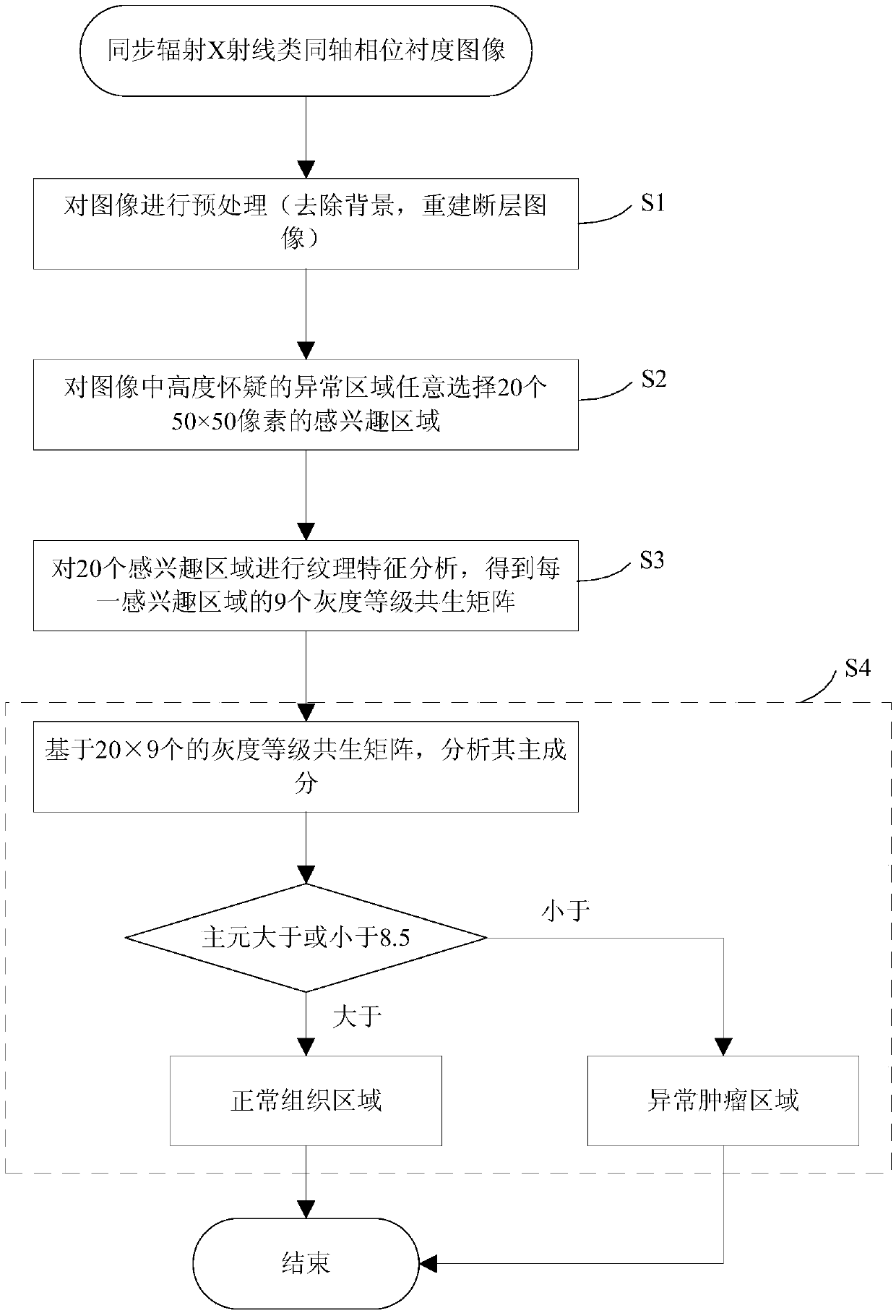
[0067] Such as figure 1 Shown is the flow chart of the tumor auxiliary diagnosis method of the present invention. The specific steps of the process are as follows:

[0068] Step 1, firstly, preprocessing the synchrotron radiation X-ray coaxial phase contrast image, including removing the background and reconstructing the tomographic image, this step is an optional step.

[0069] Step 2: Randomly select 20 regions of interest with a size of 50 × 50 pixels for highly suspected abnormal regions in the synchrotron radiation X-ray coaxial phase contrast image.

[0070] In this step, the number of selected regions of interest depends on the actual situation and is not limited to 20. The size of the region of interest depends on the actual situation and is not limited to 50×50.

[0071] Step 3: An...


PUM


Abstract

The invention discloses a tumor auxiliary diagnosis method. The tumor auxiliary diagnosis method comprises the steps of: step 1, selecting regions of interest from abnormal regions in a synchrotron radiation x ray class in-line phase contrast image; step 2, processing the regions of interest to obtain a gray level co-occurrence matrix; step 3, and carrying out principal component analysis according to the gray level co-occurrence matrix, and judging whether the abnormal regions belong to normal tissue regions or abnormal tumor regions according to analysis results.

Description

technical field [0001] The invention relates to the auxiliary diagnosis of tumors with X-ray coaxial phase contrast images in biomedical engineering, in particular to a method for auxiliary diagnosis of tumors. Background technique [0002] Synchrotron radiation X-ray coaxial phase contrast imaging is a new imaging technology, which can image various substances. In biomedical applications, synchrotron radiation X-ray coaxial phase contrast imaging can image biological tissues, and its imaging resolution is on the order of microns. Synchrotron X-ray coaxial phase-contrast imaging is currently in the experimental research stage. It has a good image resolution for biological soft tissue images, and can detect early tumors by an order of magnitude compared with existing imaging equipment. [0003] At present, the existing gray-level co-occurrence matrix texture feature method is mainly used in existing image images. For example, most of the gray-level co-occurrence matrix textu...

Claims


Application Information

Patent Timeline
IPC IPC(8): G06T7/00G06T7/40
CPCG06T7/0012G06T2207/30096
Inventor 陶蔷罗述谦
Owner CAPITAL UNIVERSITY OF MEDICAL SCIENCES
Features
  • Generate Ideas
  • Intellectual Property
  • Life Sciences
  • Materials
  • Tech Scout
  • Unparalleled Data Quality
  • Higher Quality Content
  • 60% Fewer Hallucinations
Learn More