A nanofibrous tissue filler and its preparation method

A technology of nanofibers and fillers, applied in the fields of surgery, medical science, etc., can solve the problems of easy deformation, difficult to support, and it is difficult for tissue fluid to infiltrate the stent.

Active Publication Date: 2018-12-21
MEDPRIN REGENERATIVE MEDICAL TECH
View PDF5 Cites 0 Cited by
  • Summary
  • Abstract
  • Description
  • Claims
  • Application Information

AI Technical Summary

Problems solved by technology

However, the pores of the nanofibrous scaffold prepared by this method are very small, which are micron-sized, and the middle is filled with air, which is relatively easy to deform and difficult to play a supporting role; Due to the shielding effect of air, it is difficult for tissue fluid to infiltrate the stent, and an accumulation of tissue fluid will also form around the stent

Method used


Image

  • A nanofibrous tissue filler and its preparation method
  • A nanofibrous tissue filler and its preparation method
  • A nanofibrous tissue filler and its preparation method

Examples


Embodiment 1

[0055] S1. Add 0.8g of polylactic acid into 10ml of hexafluoroisopropanol solution, stir at room temperature until dissolved, and make 8% (w / v) spinning solution; add 10g of gelatin into 100ml of deionized water, heat to 40 Stir until dissolved at ℃ to make a 10% (w / v) treatment solution; add 500ml of absolute ethanol to the crystallization dish and heat to 55℃ as a coagulation bath;

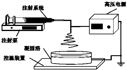
[0056] S2. If figure 1 As shown, add the spinning solution into the syringe, add an extension tube to the front end of the syringe and connect a 20G needle, place the syringe on the micro-injection pump, the needle is perpendicular to the crystallization dish, and the lower part of the crystallization dish is grounded; set the injection rate to 2ml / h, When the solution is extruded from the needle tip, a voltage of 18kv is applied to the needle tip; at this time, nanofibers are ejected and collected in the coagulation bath; when the collected nanofiber cluster reaches a certain amount, the voltag...

Embodiment 2

[0062] S1. Add 0.6g of polycaprolactone into 10ml of hexafluoroisopropanol solution, stir at room temperature until dissolved to make a 6% (w / v) spinning solution; add 1g of hyaluronic acid into 100ml of deionized water , stir until dissolved, then add 2g of antibiotic ceftriaxone sodium powder to the treatment solution to make a treatment solution with a hyaluronic acid concentration of 1% (w / v) and a ceftriaxone sodium concentration of 20mg / ml; 500ml of absolute ethanol Add it to a crystallization dish and heat it to 55°C as a coagulation bath;

[0063] S2. If figure 1 As shown, add the spinning solution into the syringe, add an extension tube to the front end of the syringe and connect a 20G needle, place the syringe on the micro-injection pump, the needle is perpendicular to the crystallization dish, and the lower part of the crystallization dish is grounded; set the injection rate to 3ml / h, When the solution is squeezed out from the needle tip, a voltage of 20kv is appli...

Embodiment 3

[0068] S1. Add 0.8g of polylactic acid into 10ml of hexafluoroisopropanol solution, stir at room temperature until dissolved, and make 8% (w / v) spinning solution; add 10g of gelatin into 100ml of deionized water, heat to 40 Stir at ℃ until dissolved, then dissolve 1g of sodium alginate in the above treatment solution, the concentration of gelatin in the final treatment solution is 10% (w / v), the concentration of sodium alginate is 1% (w / v); 5g Calcium chloride was dissolved in 95ml of deionized water to make a gel solution; 500ml of absolute ethanol was added to the crystallization dish and heated to 55°C as a coagulation bath;

[0069] S2. If figure 1 As shown, add the spinning solution into the syringe, add an extension tube to the front end of the syringe and connect a 20G needle, place the syringe on the micro-injection pump, the needle is perpendicular to the crystallization dish, and the lower part of the crystallization dish is grounded; set the injection rate to 2ml / h,...


PUM

PropertyMeasurementUnit
diameteraaaaaaaaaa
diameteraaaaaaaaaa
densityaaaaaaaaaa

Abstract

The invention discloses a nanofiber tissue filler and a preparing method thereof. The nanofiber tissue filler comprises a frame formed by nanofiber and a filling material filling among the nanofiber, the nanofiber frame is formed by the nanofiber prepared from a slow-to-degrade material, and the filling material is prepared from a fast-to-degrade material. The method comprises: using a dry spraying wet method electrospinning technique to prepare the slow-to-degrade material into a spinning solution and then performing electrospinning for coagulating bath, obtaining nanofiber agglomerates by receiving, then cleaning to remove the coagulating bath, soaking in fast-to-degrade material solution, and finally sizing, and freeze-drying to form the nanofiber tissue filler. The nanofiber tissue filler has good mechanical support performance, tissue effusion eliminating performance and tissue regeneration promoting performance, histocompatibility is good without taking out, operation is simple, pain and an economic burden of a patient caused by long term dressing change for many times are avoided, and the nanofiber tissue filler has very high clinical application value in filling and repairing of a tissue cavity.

Description

technical field [0001] The invention relates to the field of filling and tissue repair of tissue cavities and refractory wounds in the body, and more particularly relates to a nanofibrous tissue filler and a preparation method thereof. Background technique [0002] After thorough debridement of human soft tissue and tissue cavities such as tuberculosis, abscess, cyst, and tumor, as well as serious open wounds, penetrating defects, and severe wound contamination, a certain volume of soft tissue defects will be left. Dead space, sinus tract or fistula is formed. This kind of wound cannot be closed directly, and fluid accumulation and inflammation will occur repeatedly in the formed dead space. The usual method is to open the wound, stuff it with iodoform gauze or hose drainage, but this method needs to change the dressing repeatedly, which brings a double burden on the patient's spirit and economy. For deep dead space that cannot heal naturally, skin flap transplantation is r...

Claims


Application Information

Patent Timeline
Patent Type & Authority Patents(China)
IPC IPC(8): A61L31/06A61L31/04A61L31/14A61L31/16
CPCA61L31/042A61L31/043A61L31/044A61L31/046A61L31/048A61L31/06A61L31/146A61L31/148A61L31/16
Inventor 张海涛邓坤学袁玉宇
Owner MEDPRIN REGENERATIVE MEDICAL TECH
Why Eureka
Try Eureka