
An automatic segmentation method for microarray images of tumor tissue

A tumor tissue, automatic segmentation technology, applied in the field of medical image processing, to achieve the effect of reducing the amount of calculation and storage, less time consumption, and less workload

Active Publication Date: 2018-09-25
NANJING UNIV OF INFORMATION SCI & TECH
View PDF2 Cites 0 Cited by
  • Summary
  • Abstract
  • Description
  • Claims
  • Application Information

AI Technical Summary

Problems solved by technology

[0005] In order to solve the technical problems raised by the above-mentioned background technology, the present invention aims to provide an automatic segmentation method for tumor tissue microarray images, which overcomes the problems existing in the existing manual segmentation and extraction of tumor tissue with different data bits

Method used


Image

  • An automatic segmentation method for microarray images of tumor tissue
  • An automatic segmentation method for microarray images of tumor tissue
  • An automatic segmentation method for microarray images of tumor tissue

Examples


Embodiment Construction

[0033] The technical solutions of the present invention will be described in detail below in conjunction with the accompanying drawings.

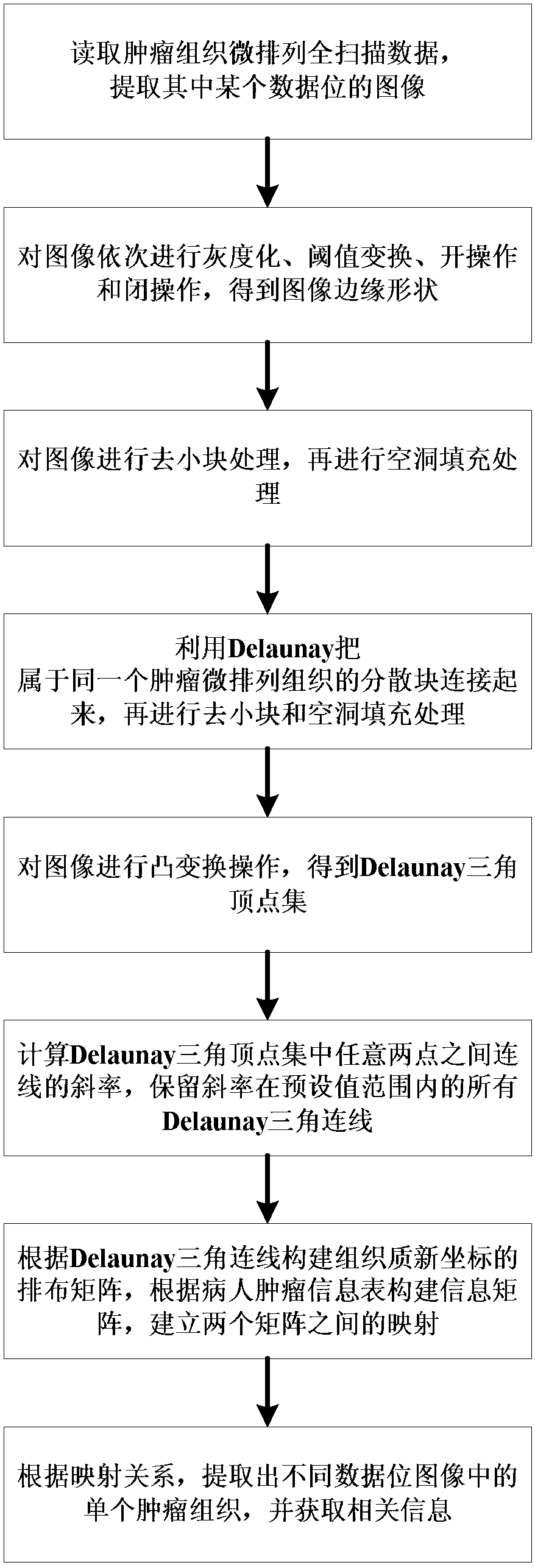
[0034] Such as figure 1 As shown, an automatic segmentation method for tumor tissue microarray images, including the following steps:

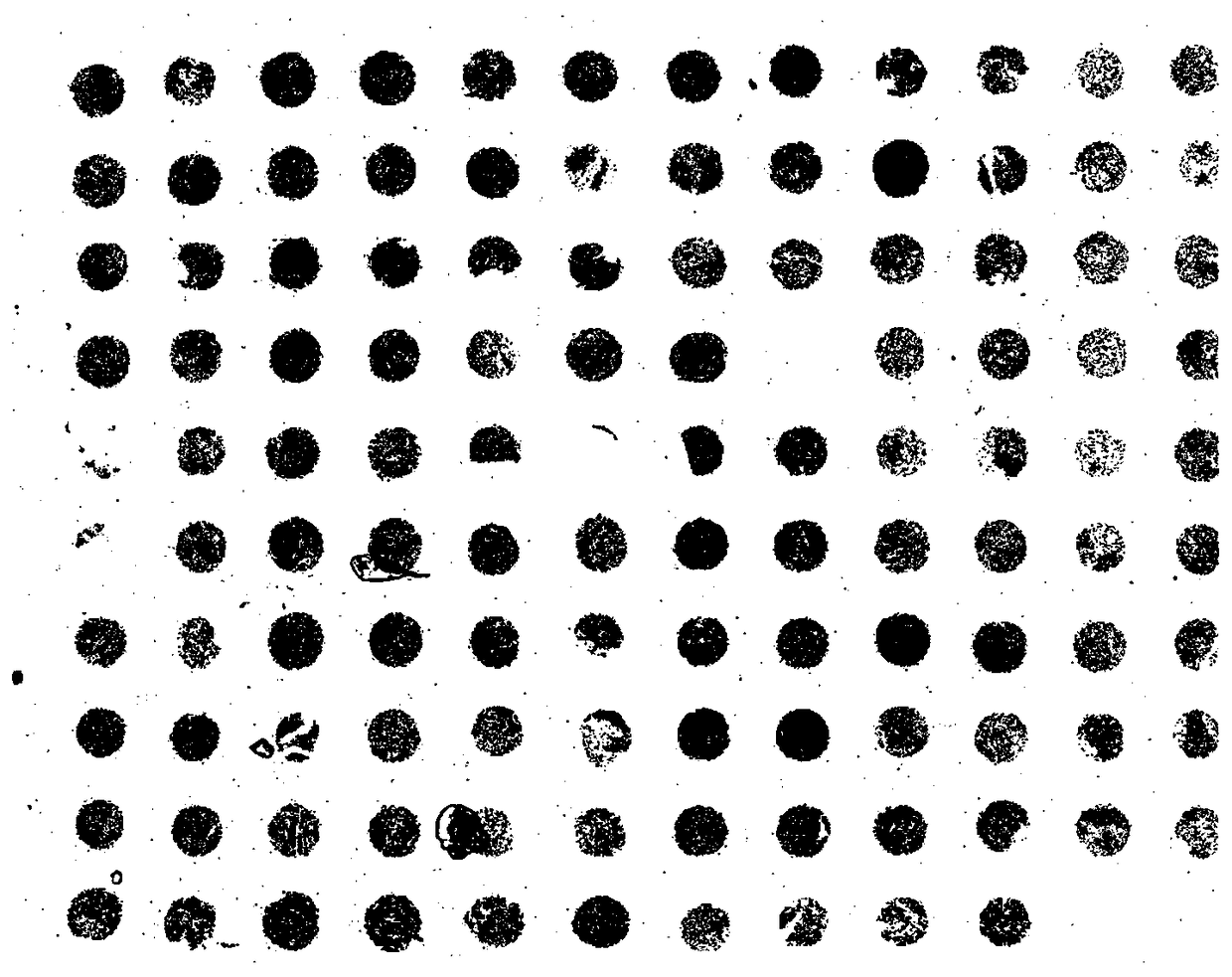
[0035] Step 1. Read the full scan data of the tumor tissue microarray, and determine how many images with different data bits are there.

[0036] Extract an image of one of the data bits.

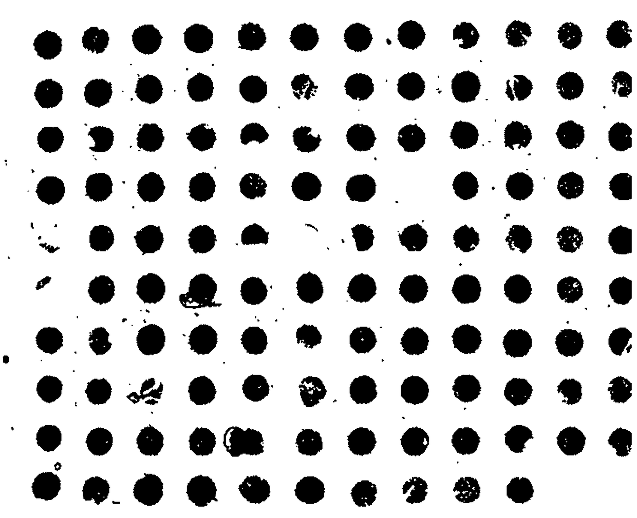
[0037] Step 2. Perform grayscale processing on the extracted image to obtain a grayscale image, convert the grayscale image into a binary image through the threshold transformation method, and then sequentially open and close the binary image to obtain a clear edge shape.

[0038] In this embodiment, the image is converted into a grayscale image by performing a weighted average method on the R, G, and B components of the image, and the conversion formula is: F(i,j)=0.30R(i,j)+0.59G( i,j)+0.11B(i,j), where F(i,j) repr...


PUM


Abstract

The invention discloses an automatic dividing method of a tumor issue micro array image. An image at a certain data bit in a pathological image is extracted. After a series of image processing steps are performed on the image, the coordinates of tumor tissues in each data thumbnail are successively found out by means of a Delaunay triangulation method. The images of individual tumor issues are successively extracted, and related information of each tumor issue is obtained. The automatic dividing method can be used for quickly extracting data at different data bits and has accuracy which is higher than that of a manual dividing solution. Furthermore the automatic dividing method has advantages of facilitating quick executing of subsequent cancer grade analysis, reducing workload in manual extraction, and furthermore reducing calculation amount and storage amount of a computer.

Description

technical field [0001] The invention belongs to the technical field of medical image processing, and in particular relates to an automatic segmentation method of tumor tissue micro-array images. Background technique [0002] With the generation of digital scanning technology for large slice images and the improvement of scanning efficiency, the digital display and storage of histopathological slides has become realistic and feasible. Higher quality analysis of pathological images is possible with digital technology. Because the characteristics of various cancers and tissues can be found from the pathological imaging images of tissue slices, and can be used to assist doctors in diagnosis, but there are still few technical researches on medical image processing. Image segmentation algorithm is very important. [0003] Medical image segmentation has a wide range of research value in medical research and practice fields such as medical research, clinical diagnosis, case analys...

Claims


Application Information

Patent Timeline
Patent Type & Authority Patents(China)
IPC IPC(8): G06T7/12G06T7/136G06T7/66
CPCG06T2207/30096
Inventor 徐军郎彬
Owner NANJING UNIV OF INFORMATION SCI & TECH
Learn More