
A Mitochondria Isolation Method Suitable for Stem Cells

A separation method and mitochondrial technology, which are applied in the field of stem cell-based biotechnology, can solve the problems of stem cells with few mitochondria and immature extraction methods, and achieve the effects of simple method, good activity and high purity.

Active Publication Date: 2020-02-18
ZHEJIANG UNIV
View PDF0 Cites 0 Cited by
  • Summary
  • Abstract
  • Description
  • Claims
  • Application Information

AI Technical Summary

Problems solved by technology

However, people mainly study the mitochondria of the main energy-consuming organs and tissues such as the heart, liver, and brain. Therefore, the methods for isolating the mitochondria of these tissues and cells are relatively mature. There are few studies on the mitochondria of stem cells, and the extraction methods are not very mature.

Method used


Image

  • A Mitochondria Isolation Method Suitable for Stem Cells
  • A Mitochondria Isolation Method Suitable for Stem Cells
  • A Mitochondria Isolation Method Suitable for Stem Cells

Examples


Embodiment 1

[0032] Example 1: Isolation of mitochondria derived from bone marrow mesenchymal stem cells.

[0033] Using the method of this example to isolate bone marrow-derived mesenchymal stem cells specifically includes the following steps:

[0034] 1) subculture amplification (P 1 -P 3 Bone marrow mesenchymal stem cells (BMSCs) were identified by flow cytometry, and their ability to differentiate into bone, cartilage and fat was identified.

[0035] 2) 2×10 7 (BMSCs), digested with 0.25% trypsin, centrifuged at 850g, 5min to collect the cells; resuspended with PBS, washed and centrifuged twice.

[0036] 3) Using the Mitochondria Isolation Kit (Mitochondria Isolation Kit, #89874, ThermoFisher Scientific), according to the operation manual, count the number of cells to determine the amount of mitochondrial extract to be prepared. Prepare mitochondrial extract solution on ice: Solution A is the lysis solution, and solution C is the termination reaction solution. Just before use, add...

Embodiment 2

[0040] Example 2: Determination of mitochondrial generation (mitochondria mass) of BMSCs cultured in vitro.

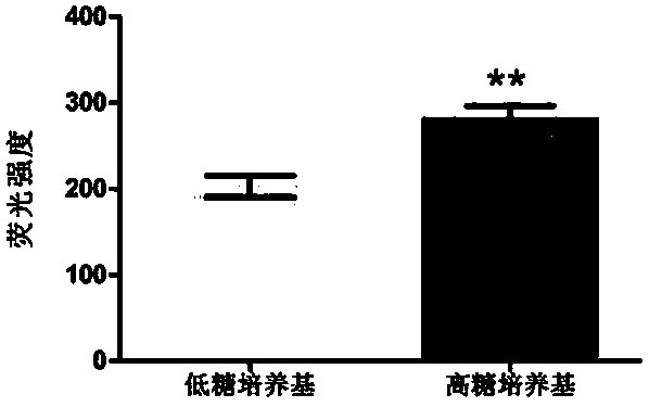
[0041] 1×10 4 BMSCs mitochondria were planted in 96-well plates (Corning) with a black bottom. A group of cells (n=42) were cultured in DMEM with 10% FBS and low glucose for 48h. Another group of cells (n=42) was cultured with 10% FBS high glucose DMEM for 48h. 200 nM MitoTrack red (Invitrogen) was then added to the medium for staining and labeling of mitochondria. First at 37°C, 5% CO 2Stain in the cell culture box for 20 minutes, then drift PSB 3 times, 10 minutes each time, and finally detect the red fluorescence intensity with a fluorescent microplate reader. The fluorescence intensity of BMSCs cultured in high-glucose medium for 48 hours was 281.3±15.03, which was significantly higher than that of BMSCs in low-glucose medium (202.7±12.37), indicating that high-glucose treatment significantly increased the generation of mitochondria.

Embodiment 3

[0042] Example 3: Determination of mitochondrial ATP content.

[0043] Using Biyuntian ATP assay kit, equilibrate the ATP assay reagent to room temperature, take 100 μl and add it to a 96-well black bottom transparent plate, then add 10 μl of freshly extracted mitochondria, shake for 30 s, let stand for 5 minutes, and immediately use the machine (Glomaxtm96MicroplateLuminometer) Determination. Quantitative determination of mitochondrial protein was performed according to the steps in the instructions of the BCA protein quantification kit. Prepare protein standards and draw a standard curve. Mix liquid A and liquid B in the kit at a ratio of 50:1, add the sample to be tested, add 2 μl of the sample to be tested, add 18 μl of PBS, and then add 200 μl of BCA incubation solution, and mix well. 37°C metal bath for 30min. Use a microplate reader with a wavelength of 562nm to measure the absorbance value of each well, and then calculate the protein concentration of the sample. Th...


PUM


Abstract

The invention provides a mitochondria isolation method suitable for stem cells. The mitochondria isolation method includes the steps of firstly, marking and identifying the detection surfaces of mesenchymal stem cells, and identifying the differentiation ability of bones, cartilage and fat; secondly, performing trypsinization, and collecting cells in a centrifugal manner; resuspending, cleaning and centrifuging; thirdly, preparing mitochondria extract which includes lysate A and reaction termination liquid C; fourthly, precipitating the cells resuspended and cleaned by the lysate A, performing vortex treatment, standing, and repeating for several times; adding the reaction termination liquid C to stop the pyrolysis, and centrifuging to obtain white precipitate for the fifth step; fifthly, repeating the fourth step on the precipitate obtained by the first centrifuging in the fourth step; sixthly, placing the white precipitate obtained in the fourth step and the white precipitate obtained in the fifth step together, and centrifuging to obtain white precipitate, and using immediately or storing at 80 DEG C. The mitochondria isolation method has the advantages that mitochondria obtained by the method is good in activity and high in purity, and the method is suitable for isolating mesenchymal stem cells to obtain the mitochondria, is simple to operate and does not need grinding.

Description

technical field [0001] The invention relates to a biotechnology based on stem cells, which is a simple method for isolating mitochondria of mesenchymal stem cells. Background technique [0002] Mitochondria is an important organelle in cells. It is unique to eukaryotic cells and exists in most living cells. It is an important organelle responsible for energy conversion. Energy substances in cells - fat, sugar, and some amino acids undergo final oxidation here, and generate ATP through coupling phosphorylation to supply the needs of cell physiological activities. Studying the structure, function and physical and chemical properties of mitochondrial inner and outer membranes, respiratory chain enzymes and mitochondrial DNA has become an important topic in cell biology research. Studies in recent years have shown that whether it is the aging process of normal cells, the canceration of tumor cells, or the proliferation and differentiation of stem cells, they are all closely rel...

Claims


Application Information

Patent Timeline
Patent Type & Authority Patents(China)
IPC IPC(8): C12N5/0775A61L27/38A61L27/54G01N33/68
CPCA61L27/3834A61L27/3895A61L27/54A61L2300/412A61L2430/00C12N5/0663G01N33/68
Inventor 王琳琳张晓明王超陈莹莹沈岳良
Owner ZHEJIANG UNIV
Learn More