
Processing device and method for processing cardiac data of a living body

A technology for processing devices and living bodies, applied in the fields of radiological diagnosis instruments, diagnosis signal processing, image data processing, etc. the effect of flexibility

Active Publication Date: 2020-02-14
KONINKLIJKE PHILIPS NV
View PDF4 Cites 0 Cited by
  • Summary
  • Abstract
  • Description
  • Claims
  • Application Information

AI Technical Summary

Problems solved by technology

This can lead to a significant increase in the time required for diagnosis or treatment

Method used


Image

  • Processing device and method for processing cardiac data of a living body
  • Processing device and method for processing cardiac data of a living body
  • Processing device and method for processing cardiac data of a living body

Examples


Embodiment Construction

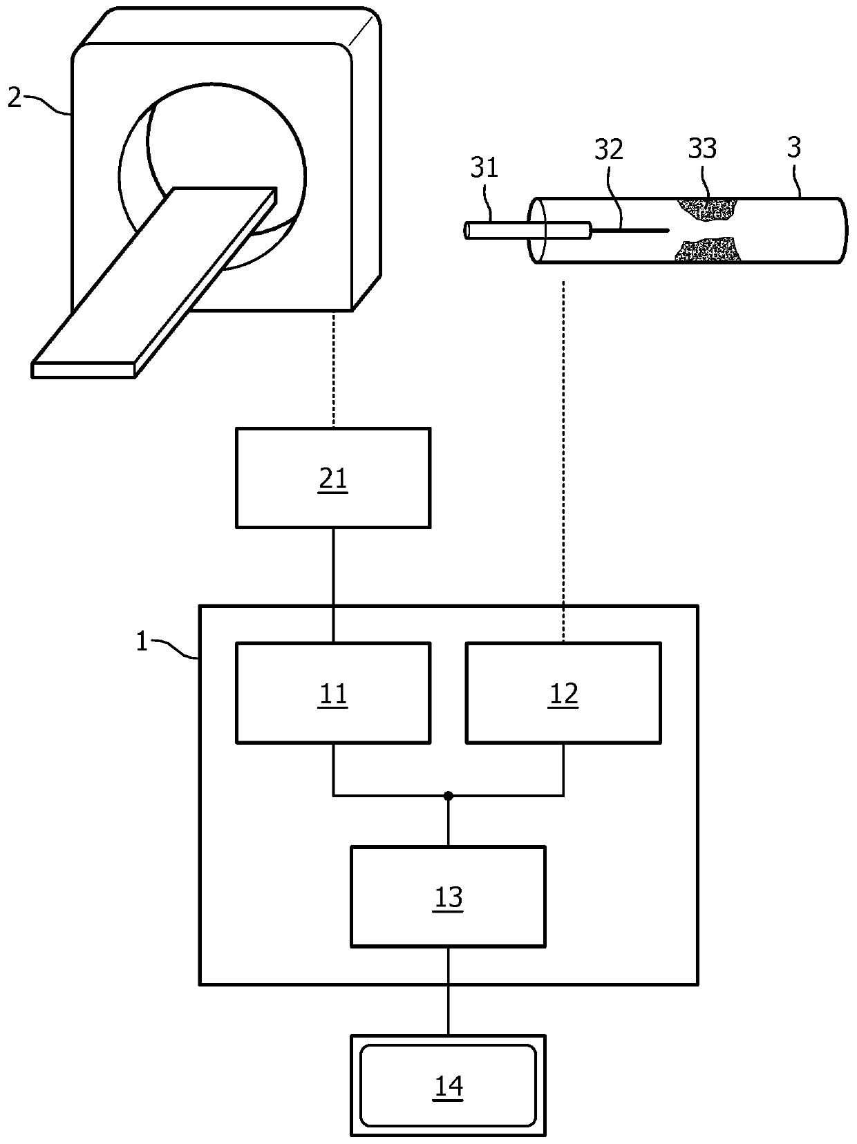
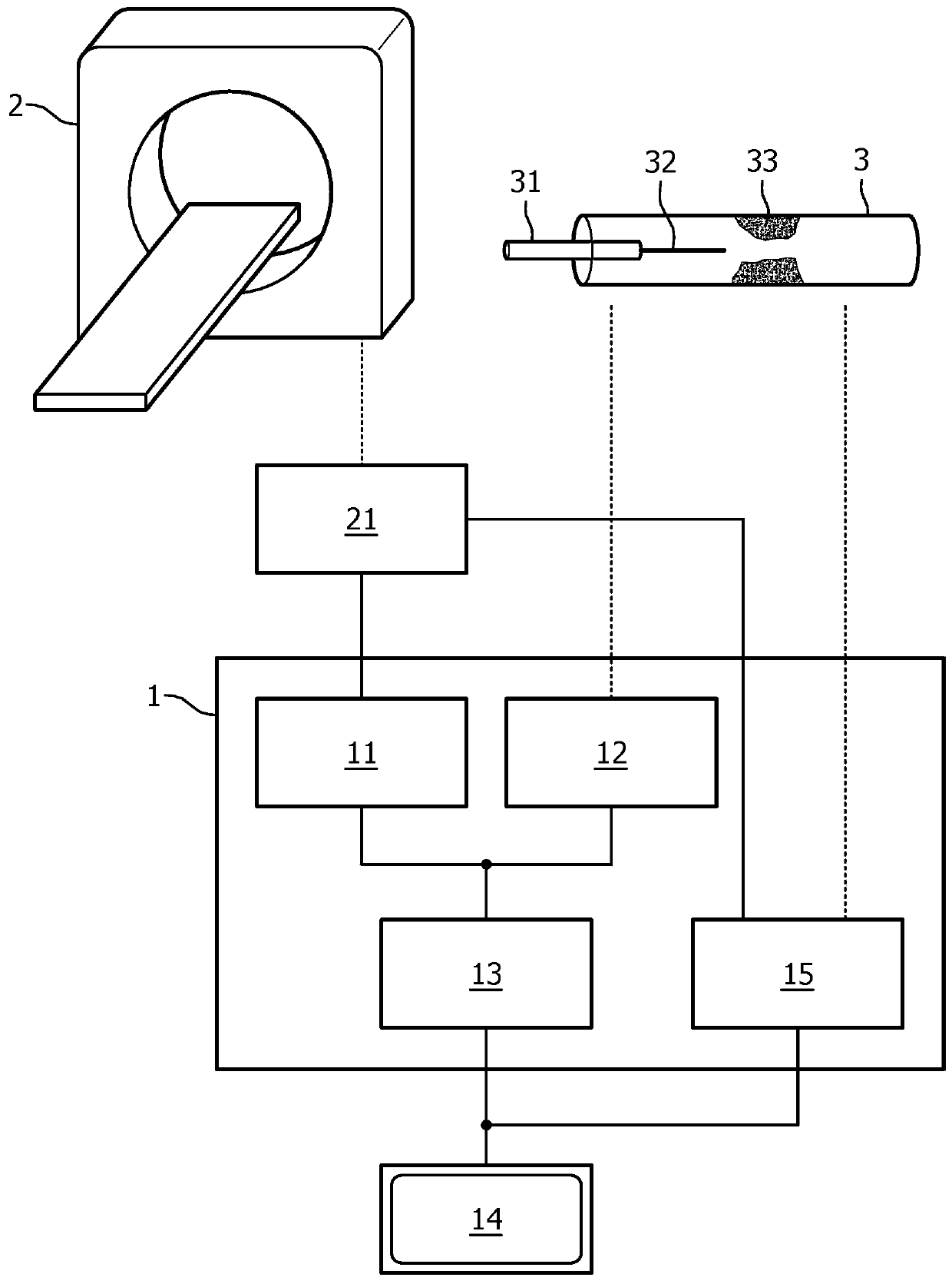
[0020] figure 1 An embodiment of a processing device according to the invention is shown schematically and by way of example. In this embodiment, the processing device 1 includes a first FFR value providing unit 11 , a second FFR value providing unit 12 , and a correction unit 13 . The FFR value is displayed by the display unit 14 .

[0021] The first FFR value providing unit 11 provides simulated FFR values ​​obtained from an image processing unit 21 which processes images obtained from a non-invasive imaging device 2 Such as computed tomography imaging equipment, ultrasound imaging equipment, positron emission tomography imaging equipment, magnetic resonance imaging equipment, X-ray imaging equipment and other non-invasive imaging equipment known to those skilled in the art or combinations thereof. The image processing unit 21 determines from the detection data detected by the non-invasive imaging device 2, for example, by calculating and simulating the first FFR value bas...


PUM


Abstract

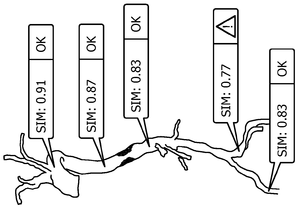
The heart data of the living body is processed by a processing unit, the processing unit comprising: a first fractional flow reserve (FFR) providing unit (11), which is used to provide a first FFR indicating the FFR of different arteries of the living body, wherein, The virtual FFR value is calculated according to the non-invasive imaging data of the artery of the living body; the second FFR providing unit (12) is used to provide the FFR value measured in the artery of the living body; the correction unit (13 ) configured to correct the first FFR value based on the second FFR value; and a display unit (14) configured to display at least the first and second FFR values ​​for corresponding locations in the coronary arteries. The first and second FFR values ​​are displayed to a cardiologist, which enables his course of action to be based on simulated and corrected values.

Description

technical field [0001] The invention relates to a processing device for processing cardiac data of a living body, a device for evaluating cardiac data of a living body, a method for evaluating stenosis in an artery of a living body, and a method for evaluating a living body A computer program product for narrowing of arteries. Background technique [0002] Degenerative stenosis is the second most common cardiovascular disease among people over 65 years old in Western Europe and North America, with an incidence rate of 2-7%, such as G.M.Feuchtner, W.Dichtl et al. in Journal of the American College of Cardiology 2006, 47( 7), as described in "Multislice Computed Tomography for Detection of Patients With Aortic Valve Stenosis and Quantification of Severity", 1410-1417. In the context of the present invention, the term stenosis denotes any abnormal narrowing of an artery. In interventional cardiology, the extent of stenosis can be measured using the Fractional Flow Reserve (FF...

Claims


Application Information

Patent Timeline
Patent Type & Authority Patents(China)
IPC IPC(8): A61B5/026
CPCA61B5/02007A61B5/026A61B5/6852A61B5/6876A61B5/72A61B6/03A61B6/504A61B6/5229A61B6/5288G06T7/0012G06T11/003G06T2207/30048G06T2207/30101
Inventor H·施米特S·普雷弗尔哈尔H·尼克基施
Owner KONINKLIJKE PHILIPS NV
Learn More