
Framework for Abnormality Detection in Multi-Contrast Brain Magnetic Resonance Data

A technology of contrast and magnetic resonance, applied in the field of abnormal detection, can solve the problem of inability to improve patient-specific test benches

Active Publication Date: 2017-03-22
SIEMENS HEALTHCARE GMBH
View PDF4 Cites 2 Cited by
  • Summary
  • Abstract
  • Description
  • Claims
  • Application Information

AI Technical Summary

Problems solved by technology

Additionally, state-of-the-art medical image analysis schemes often employ supervised learning schemes that require carefully designed features / biomarkers and large amounts of training data for "robustness"
These protocols may fail to advance patient-specific test-beds, and they are implicitly or explicitly designed to infer models or representations of abnormalities that may not be needed

Method used


Image

  • Framework for Abnormality Detection in Multi-Contrast Brain Magnetic Resonance Data
  • Framework for Abnormality Detection in Multi-Contrast Brain Magnetic Resonance Data
  • Framework for Abnormality Detection in Multi-Contrast Brain Magnetic Resonance Data

Examples


Embodiment Construction

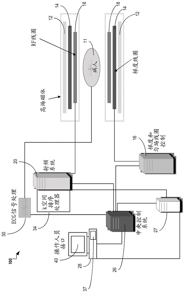
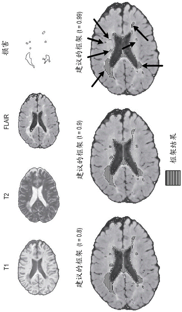
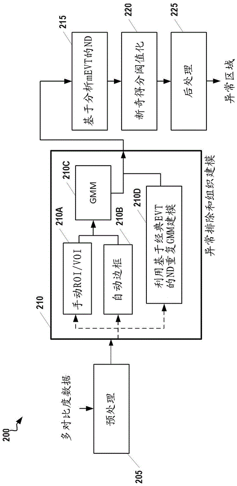
[0022] The following disclosure describes the invention in terms of several embodiments relating to methods, systems and devices related to a complete medical image analysis framework for detecting abnormalities in multi-contrast brain magnetic resonance (MR) data. More specifically, the medical image analysis framework described here accepts multi-contrast (T1 / T2 / PD / FLAIR / SWI) brain MR data of a single subject, identifies normal tissue with or without user guidance, performs these Normal tissue is parametrically modeled, and a slightly modified version of novelty detection (ND) using multivariate extreme value theory (EVT) is applied to all image data in order to detect abnormalities, if any, in the subject's brain. This framework can be applied, for example, to the detection of multiple sclerosis, traumatic brain injury, ischemic stroke, and atypical gliomas, and thus, can be used to monitor treatment.

[0023] figure 1 A system 100 for sequencing the acquisition of frequen...


PUM


Abstract

A computer-implemented method for identifying abnormalities in Magnetic Resonance (MR) brain image data includes a computer receiving multi-contrast MR image data of a subject's brain and identifying, within the multi-contrast MR image data, (i) an abnormality region comprising one or more suspected abnormalities and (ii) a healthy region comprising healthy tissue. The computer creates a model of the healthy region, computes a novelty score for each voxel in the multi-contrast MR image data based on the abnormality region and the model, and creates an abnormality map of the subject's brain based on the novelty score computed for each voxel in the multi-contrast MR image data.

Description

technical field [0001] The present invention generally relates to methods, systems and devices for detecting abnormalities in multi-contrast magnetic resonance imaging (MRI) brain data. The disclosed techniques can be applied, for example, to the detection of multiple sclerosis (MS), traumatic brain injury, ischemic stroke, and atypical glioma. Background technique [0002] The problem of automatically detecting abnormalities in imaging data, eg pathology in the form of tumors, lesions or structures such as metallic implants, has been a topic of interest for several years. In particular, since the advent of current multi-contrast (e.g., T1 / T2 / PD / FLAIR / SWI) MR imaging protocols, diffuse abnormal lesions (e.g., encountered in brain images of patients with multiple sclerosis, traumatic brain injury Advances have been made in the detection and delineation of hyperintensity obtained. However, this problem is challenging due to the complex representation of pathology (eg, highly...

Claims


Application Information

Patent Timeline
Patent Type & Authority Applications(China)
IPC IPC(8): A61B5/055
CPCA61B5/0042A61B5/055A61B5/72A61B2576/026A61B5/7267A61B5/0013A61B5/0037A61B5/08A61B5/201A61B5/4064G16H50/50
Inventor H.E.策廷古尔M.S.纳达尔B.L.奥德里
Owner SIEMENS HEALTHCARE GMBH
Learn More