Deep model and shallow model decision fusion-based pulmonary nodule CT image automatic classification method

A technology of decision fusion and CT images, applied in the fields of image processing and medical integration, can solve problems such as differential classification performance, and achieve the effect of avoiding changes in feature space distribution and overcoming poor results

Inactive Publication Date: 2017-05-10
NORTHWESTERN POLYTECHNICAL UNIV
View PDF4 Cites 59 Cited by
  • Summary
  • Abstract
  • Description
  • Claims
  • Application Information

AI Technical Summary

Problems solved by technology

In the present invention, when we perform feature layer fusion, a feature may be affected by other features, and then the spatial distribution of these features will change, and these changes will lead to poor classification performance

Method used


Image

  • Deep model and shallow model decision fusion-based pulmonary nodule CT image automatic classification method
  • Deep model and shallow model decision fusion-based pulmonary nodule CT image automatic classification method
  • Deep model and shallow model decision fusion-based pulmonary nodule CT image automatic classification method

Examples


Embodiment Construction

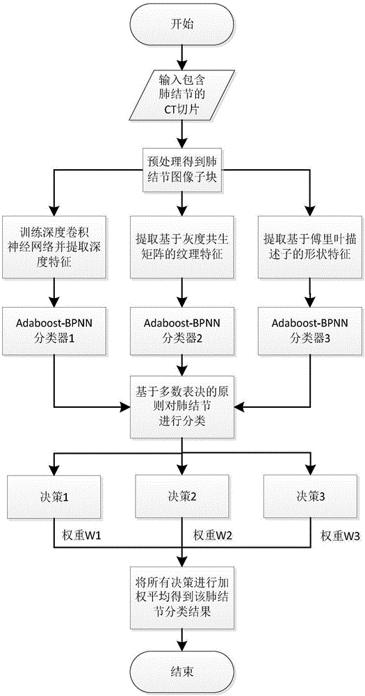
[0045]The invention provides a weight-based multi-feature decision-making classification algorithm. This method extracts image subblocks that just contain pulmonary nodules from each CT slice image containing pulmonary nodules, and then unifies the image subblocks of different sizes to a size of 32×32. Since pulmonary nodules are three-dimensional spheroids, a complete CT image of pulmonary nodules contains multiple slices, and the category of each slice is the category of its CT image, so the pulmonary nodule classification problem based on three-dimensional CT images Converted to a classification problem on two-dimensional space. Next, feature extraction is carried out. First, the stochastic gradient descent method is used to train the deep convolutional neural network model on all preprocessed training image blocks, and the output of the fully connected layer of the network is selected as the description feature of the corresponding image block, which is called It is a dep...


PUM


Abstract

The invention relates to a deep model and shallow model decision fusion-based pulmonary nodule CT image automatic classification method. Deep convolutional neural network-based features and visual features describing pulmonary nodule textures and shapes are extracted. Three classifiers are trained for the three different features, and results of all the classifiers are subjected to weighted averaging to obtain a final classification result. Therefore, the innovation of a CT image-based pulmonary nodule classification method is realized.

Description

technical field [0001] The invention belongs to the field of image processing and medical integration, and specifically relates to a method for classifying benign and malignant pulmonary nodules based on CT images. We extract features based on deep convolutional neural networks and visual features describing the texture and shape of pulmonary nodules. Three classifiers are trained for these three different features, and the results of all classifiers are weighted and averaged to obtain the final classification result, which realizes the innovation of the lung nodule classification method based on CT images. Background technique [0002] Classification of pulmonary nodules based on CT images can generally be divided into three parts: pulmonary nodule segmentation, feature extraction, and classification of pulmonary nodules. Pulmonary nodule segmentation is based on the coordinates of the nodules marked by experts, and the nodules at the corresponding positions are segmented i...

Claims


Application Information

Patent Timeline
Patent Type & Authority Applications(China)
IPC IPC(8): G06K9/62G06N3/04G06N3/08
CPCG06N3/08G06N3/045G06F18/2413
Inventor 夏勇谢雨彤张艳宁
Owner NORTHWESTERN POLYTECHNICAL UNIV
Why Eureka
Try Eureka