Ultrasonic 3d fetal facial contour image processing method and system

An image processing and fetal technology, applied in ultrasonic/sonic/infrasonic diagnosis, sonic diagnosis, infrasonic diagnosis, etc., can solve the problems of long time, complicated operation, difficult target observation, etc., to simplify the operation and improve the drawing rate. Effect

Active Publication Date: 2017-05-31
SONOSCAPE MEDICAL CORP
View PDF6 Cites 11 Cited by
  • Summary
  • Abstract
  • Description
  • Claims
  • Application Information

AI Technical Summary

Problems solved by technology

However, due to the particularity of the imaging environment, when performing 3D visualization of the fetal face, the front of the fetal face may be blocked by the placenta, umbilical cord, arms, uterine wall, etc., so that the collected 3D volume data may include placenta, amniotic fluid suspension Substances, umbilical cords, uterine tissues, etc. will block the imaging target and make it difficult to observe the target
[0003] Although there is usually an interactive volume cutting method on the current ultrasonic inspection equipment that can provide the inspector to cut the occluded part, but the operation is cumbersome and time-consuming

Method used


Image

  • Ultrasonic 3d fetal facial contour image processing method and system
  • Ultrasonic 3d fetal facial contour image processing method and system
  • Ultrasonic 3d fetal facial contour image processing method and system

Examples


Embodiment Construction

[0059] In order to make the technical problems solved by the present invention, the technical solutions adopted and the technical effects achieved clearer, the technical solutions of the embodiments of the present invention will be further described in detail below in conjunction with the accompanying drawings. Obviously, the described embodiments are only the technical solutions of the present invention. Some, but not all, embodiments. Based on the embodiments of the present invention, all other embodiments obtained by those skilled in the art without creative efforts fall within the protection scope of the present invention.

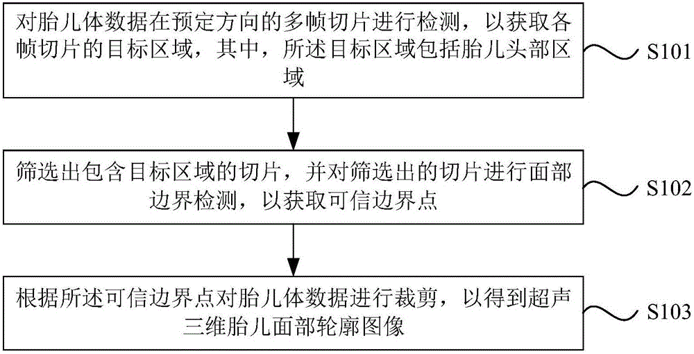
[0060] Please refer to figure 1 , which is a flow chart of the ultrasonic three-dimensional fetal facial contour image processing method provided in the specific embodiment of the present invention. Such as figure 1 As shown, the method includes:

[0061] Step S101 : Detect multiple frames of slices of fetal volume data in a predetermined direction ...


PUM


Abstract

The invention discloses an ultrasonic 3d fetal facial contour image processing method and system. The method comprises the steps that multiple frames of slices of the fetal body data in the preset direction are tested and the objective area of each frame of slice is obtained, wherein the objective area comprises a fetal head area; the slices of the objective area are screened out, and the facial boundary tests are performed on the selected slices so as to obtain a credible boundary point; the fetal body data is clipped according to the credible point to obtain the ultrasonic 3d facial contour image. The system uses credible boundary point clipping data and automatically clips the shielding part of the fetal face, so that the test human operation is simplified and the ultrasonic 3d plotting rate is increased.

Description

technical field [0001] The invention relates to the technical field of ultrasonic imaging, in particular to an ultrasonic three-dimensional fetal facial contour image processing method and system. Background technique [0002] The 3D ultrasound imaging system is based on the traditional 2D ultrasound, which collects multiple sequential spatial sequences of 2D ultrasound images, and reproduces the volumetric data through steps such as scan conversion according to the spatial position relationship of data collection. The three-dimensional ultrasound imaging system provides high-level one-dimensional spatial information that cannot be provided by traditional two-dimensional ultrasound, making clinical diagnosis and observation more intuitive and flexible, and smoother communication between doctors and patients on diagnosis results. Because of its rich and intuitive information, the 3D ultrasound imaging system is currently mainly used for observation of fetal morphology in the ...

Claims


Application Information

Patent Timeline
Patent Type & Authority Applications(China)
IPC IPC(8): A61B8/00
CPCA61B8/5215
Inventor 黄柳倩孙慧艾金钦刘旭江喻美媛
Owner SONOSCAPE MEDICAL CORP
Why Eureka
Try Eureka