

A method and device for processing liver scan images
A processing method and image technology, applied in the field of image processing, can solve the problems of insufficient stability, objectivity and incomplete reliability of the analysis method, and achieve stable and objective results, improve reliability, and avoid interference effects.
- Summary
- Abstract
- Description
- Claims
- Application Information
AI Technical Summary
Problems solved by technology
Method used
Image

Examples
Embodiment 1
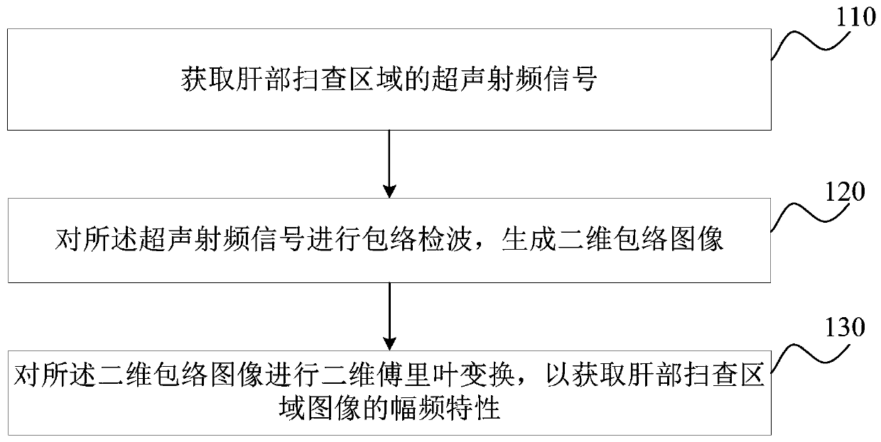
[0049] figure 1 It is a schematic flow chart of the method for processing liver scan images provided by Embodiment 1 of the present invention. This embodiment is applicable to the case of obtaining liver fibrosis degree parameters, and this method can be executed by a processing device for liver scan images. The device can be realized by software / hardware, and can be integrated into the ultrasonic imaging equipment.
[0050] see figure 1 , the processing method of the scan image of the liver, comprising:
[0051] S110. Acquire ultrasonic radio frequency signals of the scan region of the liver.
[0052] During ultrasound scanning, the ultrasound radio frequency signal is processed through a traditional B-mode imaging process to obtain a two-dimensional liver image. The acquired two-dimensional liver image at this time is generated based on the echo signal. All ultrasonic radio frequency signals corresponding to the region are determined according to the scanned region of th...
Embodiment 2
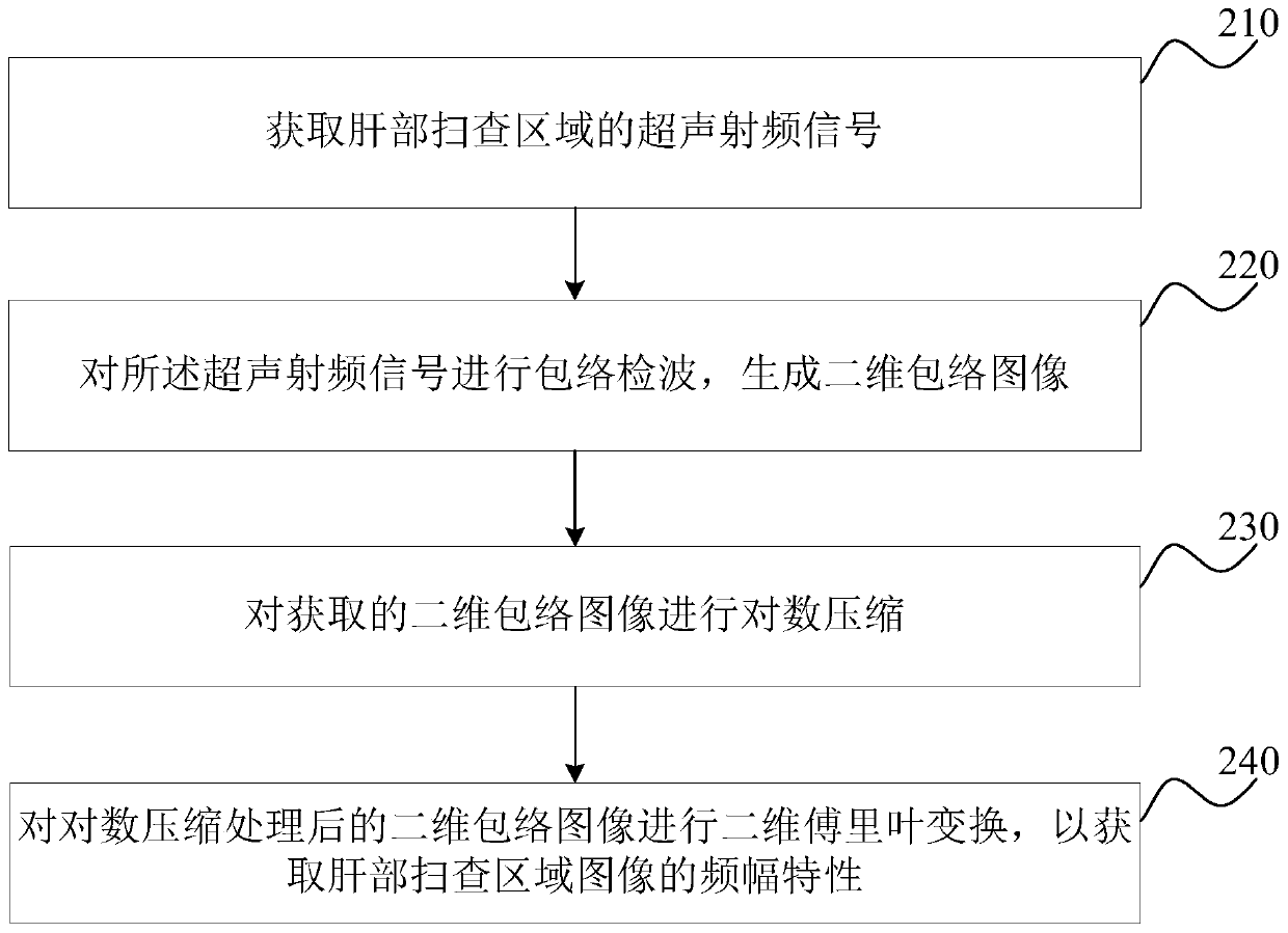
[0060] figure 2 It is a schematic flowchart of a method for processing liver scan images provided in Embodiment 2 of the present invention. This embodiment is optimized on the basis of the above embodiments. In this embodiment, after acquiring the two-dimensional envelope image, before performing two-dimensional Fourier transform on the two-dimensional envelope image, the following steps are added: Perform logarithmic compression on the two-dimensional envelope image; and carry out two-dimensional Fourier transform on the two-dimensional envelope image, specifically optimized as: perform two-dimensional compression on the two-dimensional envelope image after logarithmic compression Fourier transform.
[0061] The method for processing liver scan images provided in this embodiment specifically includes:
[0062] S210. Acquire ultrasonic radio frequency signals of the scan region of the liver.
[0063] S220. Perform envelope detection on the ultrasonic radio frequency signal...
Embodiment 3
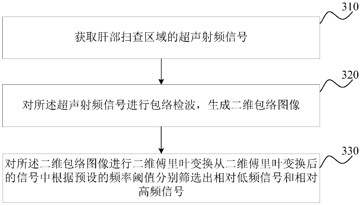
[0070] image 3 It is a schematic flowchart of the processing method of the liver scan image provided by the third embodiment of the present invention. This embodiment is optimized on the basis of the above embodiments. In this embodiment, the two-dimensional Fourier transform is performed on the two-dimensional envelope image to obtain the amplitude-frequency characteristics of the image of the liver scan area. , the specific optimization is as follows: from the two-dimensional Fourier transformed signal, the relatively low-frequency signal and the relatively high-frequency signal are respectively screened out according to a preset frequency threshold.
[0071] The method for processing liver scan images provided in this embodiment specifically includes:
[0072] S210. Acquire ultrasonic radio frequency signals of the scan region of the liver.
[0073] S220. Perform envelope detection on the ultrasonic radio frequency signal to generate a two-dimensional envelope image.
...
PUM
 Abstract
Description
Claims
Application Information