A Localization Method of Renal Cortex Based on Statistical Shape Model

A statistical shape and positioning method technology, applied in the field of medical imaging algorithms, can solve the problems of poor recognition and positioning of renal cortex, difficult to distinguish, and low efficiency of model training

Active Publication Date: 2019-11-29
SUZHOU UNIV
View PDF2 Cites 0 Cited by
  • Summary
  • Abstract
  • Description
  • Claims
  • Application Information

AI Technical Summary

Problems solved by technology

[0003] The existing medical imaging technology can not do a good job of identifying and positioning the renal cortex. The main reason is that the kidney is different from other organs such as the liver, and the anatomical structure of the kidney is relatively complicated.
The kidney has four anatomical structures, among which the renal cortex and renal column are connected, and the renal cortex and renal column have similar reflection intensity to light, so it is difficult to distinguish in medical imaging
In addition, the kidney and adjacent organs, such as the liver and spleen, often overlap in the image, so it is difficult to accurately model in medical imaging
Existing localization methods based on learning algorithms cannot accurately localize the renal cortex, and the model training efficiency is not high

Method used


Image

  • A Localization Method of Renal Cortex Based on Statistical Shape Model
  • A Localization Method of Renal Cortex Based on Statistical Shape Model
  • A Localization Method of Renal Cortex Based on Statistical Shape Model

Examples


Embodiment

[0051] Embodiment: The renal cortex localization method of the present invention is based on the statistical shape model of the renal cortex, and is intended to make full and reasonable use of the statistical shape information of the renal cortex in the image for localization. Let's take a CT image as an example. The positioning method is divided into training phase and testing phase, as follows:

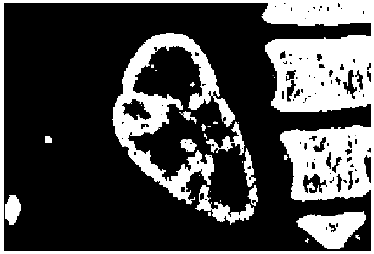
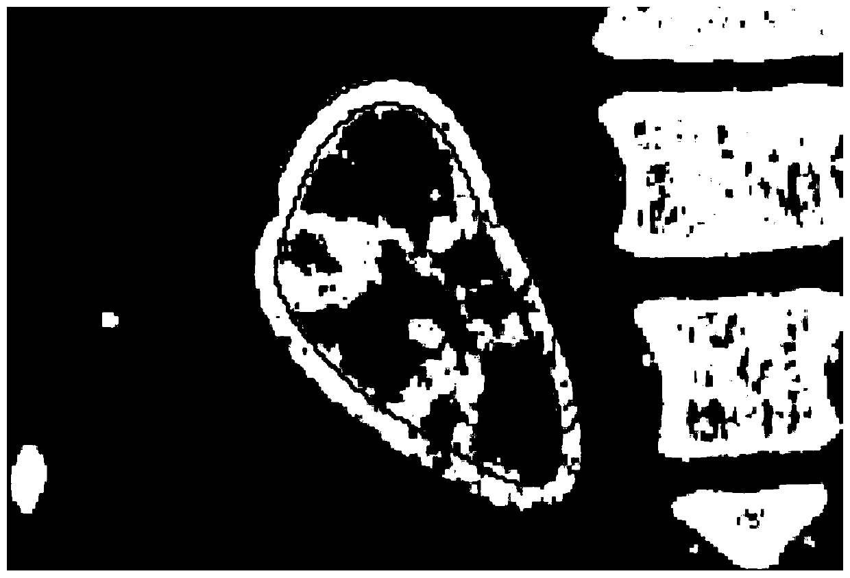
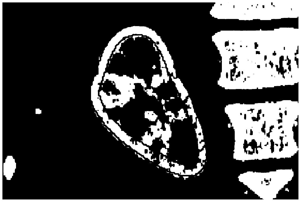
[0052] During the training phase, kidneys are manually labeled for each 3D CT image in the training dataset. Such as figure 1 It is a slice image of abdominal CT. Label the renal column, renal medulla and other structures in the kidney as the same type of L1, such as figure 2 The area contained in the (grayscale) inner circle in , marks the whole kidney as another type of L2, such as figure 2 The area enclosed by the (grayscale) outer circle in .

[0053] 2. Use the marching cube algorithm to convert the binary data in the marked areas of L1 and L2 into surface data M1 and M2...


PUM


Abstract

The invention discloses a renal cortex locating method based on a statistical shape model. The method includes a training stage and a renal cortex locating stage. The method is characterized in that in the training stage, artificial marks L1 and L2 of a kidney of each three-dimensional CT image in a training data set are made, binary data in L1 and L2 marking regions is converted to surface data M1 and M2 in a corresponding manner, interior surface data of renal cortex is calculated, and a renal cortex statistical shape model is established. According to the method, in the training stage, the renal cortex statistical shape model is established to perform statistics on the change mode of renal cortex, and the accuracy and the rapidity of locating of renal cortex are improved by employing an iteration model deformation algorithm.

Description

technical field [0001] The invention belongs to the field of medical imaging algorithms, and in particular relates to a renal cortex positioning method based on a statistical shape model. Background technique [0002] The renal cortex is an important part of the kidney. About 1.9% of adults are diagnosed with visceral diseases, and a considerable number of them die from nephritis, nephrotic syndrome, nephropathy and other diseases related to the renal cortex. Therefore The diagnostic research on the renal cortex is of great significance in the study of renal diseases. [0003] The existing medical imaging technology can not do a good job of identifying and positioning the renal cortex. The main reason is that the kidney is different from other organs such as the liver, and the anatomical structure of the kidney is relatively complicated. The kidney has four anatomical structures, among which the renal cortex and renal column are connected, and the renal cortex and renal col...

Claims


Application Information

Patent Timeline
Patent Type & Authority Patents(China)
IPC IPC(8): G06T7/00G06T7/73G06T7/11G06T7/136G06T7/66
CPCG06T7/0012G06T2207/10081G06T2207/20081G06T2207/30084
Inventor 向德辉陈新建
Owner SUZHOU UNIV
Why Eureka
Try Eureka