Device and method for collecting and segmenting of hyperspectral images of unstainedpathological sections

A technology of hyperspectral images and pathological slices, which is applied in the field of devices for rapid acquisition and segmentation of non-stained pathological slices, and can solve time-consuming problems

Active Publication Date: 2017-08-18
XI AN JIAOTONG UNIV
View PDF5 Cites 27 Cited by
  • Summary
  • Abstract
  • Description
  • Claims
  • Application Information

AI Technical Summary

Problems solved by technology

However, both of these two methods require staining of cancer sections, which still requi

Method used


Image

  • Device and method for collecting and segmenting of hyperspectral images of unstainedpathological sections
  • Device and method for collecting and segmenting of hyperspectral images of unstainedpathological sections
  • Device and method for collecting and segmenting of hyperspectral images of unstainedpathological sections

Examples


Embodiment Construction

[0060] The present invention will be further described below in conjunction with the accompanying drawings.

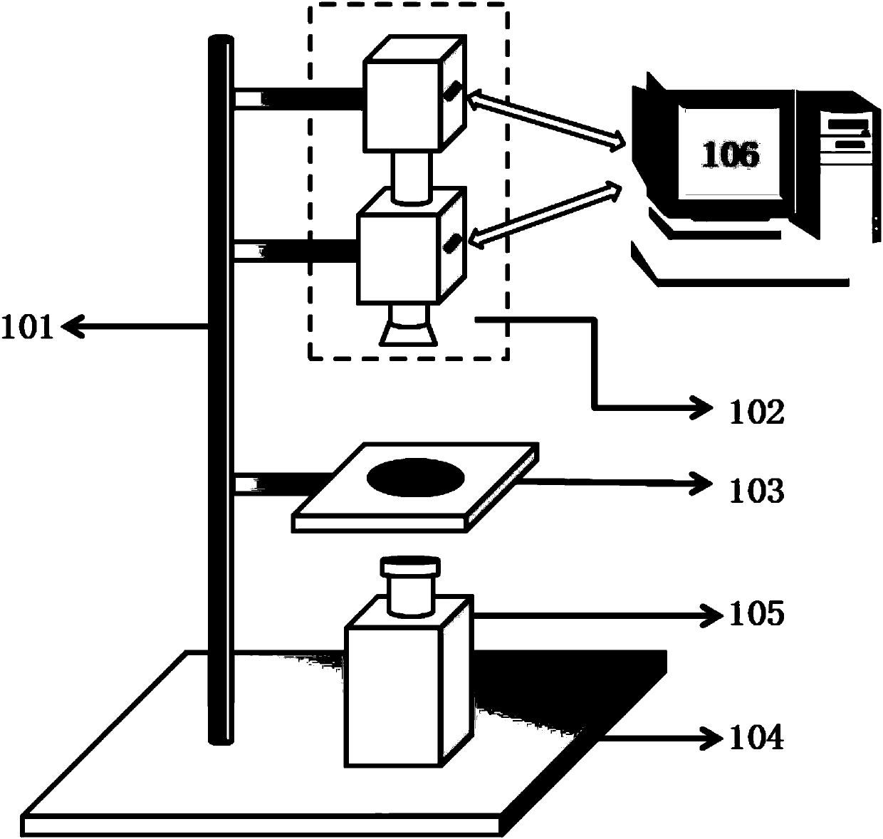
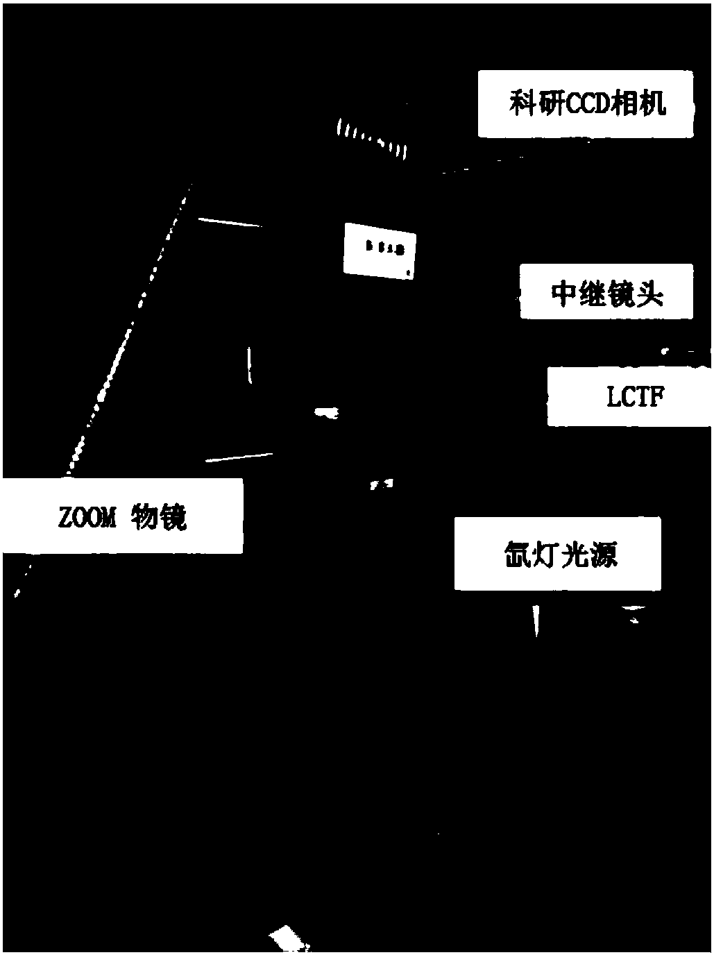
[0061] see Figure 1 to Figure 3 , a device for hyperspectral image acquisition and segmentation of non-stained pathological slices, including an alloy steel substrate 104, a xenon lamp light source 105 is placed on the alloy steel substrate 104, and a fixed bracket 101 is vertically arranged on the alloy steel substrate 104, and the fixed bracket 101 is installed from the A hyperspectral image acquisition module 102 and a sample platform 103 are arranged from top to bottom, the hyperspectral image acquisition module 102, the sample platform 103 are coaxial with the xenon lamp light source 105, and the hyperspectral image acquisition module 102 is externally connected to a computer 106;

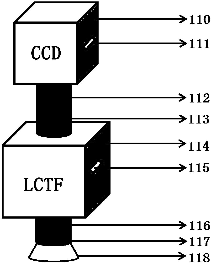
[0062] Described hyperspectral image acquisition module 102 comprises CCD camera 110, connects computer 106 through CCD camera 110 side USB interface, and CCD camera 110 is connected ...


PUM


Abstract

The invention discloses a device and method for collecting and segmenting of hyperspectral images of unstainedpathological sections. A section sample platform is supported in the middle of a bracket, the hyperspectral images of the unstainedpathological sectionsare automatically collected and processed through a computer, and lesion area segmentation resultsare obtained. Based on spectral difference caused by pathological changes of tissues, a personal computeris adopted to synchronously control relevant modules, spectral sequence images of the unstainedhistopathological sectionsare collected and preprocessed, and corresponding three-dimensionalhyperspectral dataare produced after overlaying;based the data and the combination with a currentlypopularneural network classificationthought, a spectral classification algorithm is developed to perform identity partitioning of a lesion area, thereby acceleratingthe identification rate and efficiency of the histopathological section, avoidingmanual errors which maybe brought during staining, and reducingthe time required by section making;automatic judgment is performed by utilizing a machine algorithm, therebyreducingsubjectivity caused by manual judgment. Therefore,a good auxiliary function is provided for a pathology doctor to detect the pathological sections.

Description

technical field [0001] The invention belongs to the field of detection devices, and in particular relates to a device and method for rapid collection and segmentation of non-stained pathological sections based on hyperspectral data. Background technique [0002] Pathological examination has been widely used in clinical work and scientific research. In the clinical aspect, the postmortem pathological examination and surgical pathological examination are mainly carried out. The purpose of surgical pathological examination is to clarify the diagnosis and verify the preoperative diagnosis and improve the level of clinical diagnosis; secondly, after the diagnosis is confirmed, the next treatment plan can be determined and the prognosis can be estimated, so as to improve the level of clinical treatment. Through clinical and pathological analysis, a large amount of extremely valuable scientific research data can also be obtained. [0003] A pathological slide is a type of patholo...

Claims


Application Information

Patent Timeline
IPC IPC(8): G01N21/25G01N21/01
CPCG01N21/01G01N21/25G01N2021/0181
Inventor 张镇西王森豪王晶陈韵竹张璐薇姚翠萍王斯佳
Owner XI AN JIAOTONG UNIV
Why Eureka
Try Eureka