A rib visualization aided diagnosis method for rib fractures

A technology for auxiliary diagnosis and rib fractures, applied in the field of image processing, can solve the problems of not being able to see density information, low-density tissues and organs are easily affected by missing noise, and surface shadow display methods are prone to produce artifacts, so as to improve the intuitiveness Effect

Active Publication Date: 2021-05-14
孔德兴
View PDF2 Cites 0 Cited by
  • Summary
  • Abstract
  • Description
  • Claims
  • Application Information

AI Technical Summary

Problems solved by technology

The multi-level reconstruction method requires a series of images and non-target organs are deformed; the maximum density projection method is easy to miss low-density tissues and organs and has a large impact of noise; the surface shadow display method is prone to artifacts and cannot see density information

Method used


Image

  • A rib visualization aided diagnosis method for rib fractures
  • A rib visualization aided diagnosis method for rib fractures
  • A rib visualization aided diagnosis method for rib fractures

Examples


Embodiment Construction

[0072] Below in conjunction with accompanying drawing and specific embodiment the present invention is described in further detail:

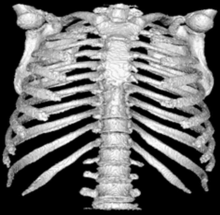
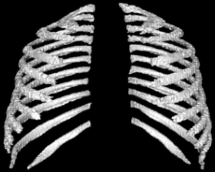
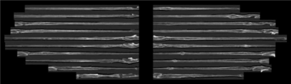
[0073] A new rib visualization aided diagnosis method for rib fractures, including the following process:

[0074] 1. Get pictures;

[0075] 2. Extract the ribs;

[0076] 3. Manual correction, removing the spine and sternum;

[0077] 4. Expand the ribs.

[0078] Said process one specifically includes: obtaining a CT scan picture of a human chest by contacting a hospital.

[0079] Described process two specifically includes:

[0080] The processing of abdominal CT images is based on the relatively high density of bones. In CT scans, the CT value is relatively large, which shows that the brightness of bones is higher than that of other tissues and organs, so there is a clear difference. By selecting an appropriate window width and window level to use The multi-scale enhancement filter operator extracts the ribs, and avoids the interference of...


PUM


Abstract

The invention relates to image processing technology and aims to provide a rib fracture auxiliary diagnosis method for rib visualization. The rib visualization aided diagnosis method for rib fractures includes the following processes: acquiring pictures; extracting ribs; manually correcting, removing the spine and sternum; and expanding the ribs. The present invention eliminates the influence of rib bending and occlusion on the diagnosis by projecting the ribs onto the plane. Compared with the traditional clinical diagnosis method, observation on the plane improves the intuitiveness of diagnosis. The present invention can be used as an aid in the diagnosis of micro fractures and fractures method.

Description

technical field [0001] The invention relates to the technical field of image processing, in particular to a rib fracture aided diagnosis method for rib visualization. Background technique [0002] Computed tomography (CT) is the main method for diagnosing rib fractures. However, due to the special organizational structure of the ribs, the ribs are obscured, so small fractures and fractures are easily missed. Accurate diagnosis requires the accumulation of experience. The conventional two-dimensional method requires manual tracking of multiple Slice, the traditional 3D visualization method needs to rotate the image during diagnosis to avoid the influence of rib occlusion. The commonly used reconstruction methods are multi-level reconstruction method, maximum density projection method and surface shadow display method. The multi-level reconstruction method requires a series of images and non-target organs are deformed; the maximum density projection method is easy to miss low...

Claims


Application Information

Patent Timeline
Patent Type & Authority Patents(China)
IPC IPC(8): G06T7/00
CPCG06T7/0012G06T2207/10081G06T2207/30008
Inventor 孔德兴严子旭陈仁栋
Owner 孔德兴
Why Eureka
Try Eureka