Heart ultrasonography bed

An ultrasound examination bed and heart technology, applied in the directions of ultrasound/sonic/infrasonic diagnosis, sonic diagnosis, infrasound diagnosis, etc., can solve the problem that the left anterior axillary line cannot be exposed, and achieve the effect of improving the inspection efficiency and reducing the burden.

Inactive Publication Date: 2017-09-19
ZHONGSHAN HOSPITAL FUDAN UNIV
View PDF4 Cites 2 Cited by
  • Summary
  • Abstract
  • Description
  • Claims
  • Application Information

AI Technical Summary

Problems solved by technology

[0004] The technical problem solved by the present invention is that in some special cases, it is necessary to place the ultrasonic probe on the left anterior axillary line to collect images, and when the examinee lies on the ordinary examination bed in the left decubitus position, the left anterior axillary line cannot be exposed. Requires patient to change position

Method used


Image

  • Heart ultrasonography bed
  • Heart ultrasonography bed
  • Heart ultrasonography bed

Examples


Embodiment Construction

[0026] The present invention will be further described below in conjunction with the accompanying drawings and specific embodiments, but not as a limitation of the present invention.

[0027] In addition, the terms "first" and "second" are used for descriptive purposes only, and cannot be interpreted as indicating or implying relative importance or implicitly specifying the quantity of indicated technical features. Thus, a feature defined as "first" and "second" may explicitly or implicitly include one or more of these features. In the description of the present invention, "plurality" means two or more, unless otherwise specifically defined.

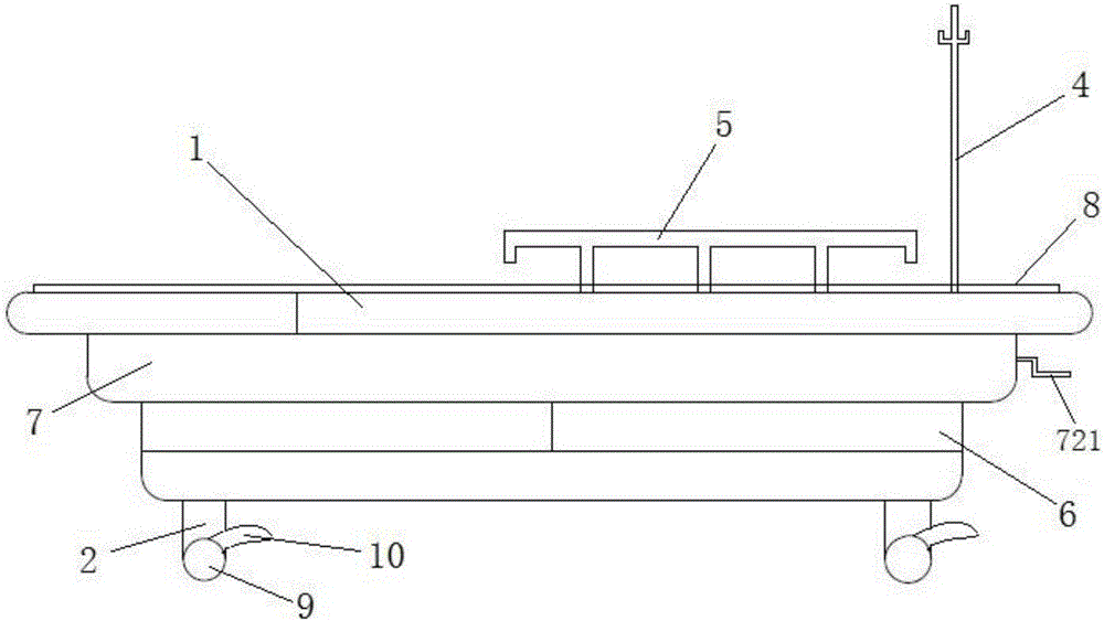
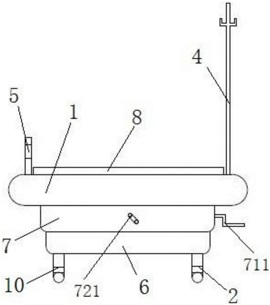
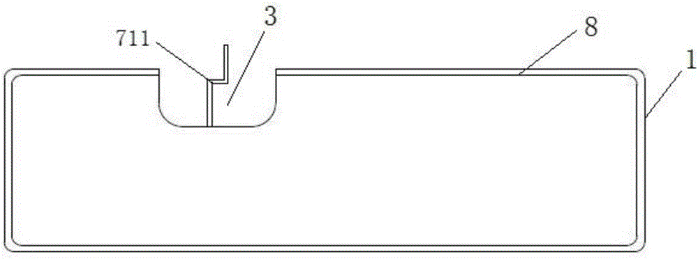
[0028] See figure 1 , figure 2 and image 3 As shown, an embodiment of the cardiac ultrasound examination bed is shown, including a bed body 1 and a bed foot 2, wherein a notch 3 is opened on the side of the bed body 1, and the notch 3 is in a semicircular structure. When the ultrasound probe needs to be placed on the left anterior ...


PUM


Abstract

The technical scheme of the invention discloses a heart ultrasonography bed. The heart ultrasonography bed comprises a bed body and bed legs, a semicircular gap is arranged at the lateral of the bed body, and the heart ultrasonography bed further comprises an infusion frame detachably installed at the lateral of the bed body and a railing reversibly installed at the edge of the bed body, and an object storage part is arranged below the bed body; a bed body adjusting mechanism is installed at the bottom of the bed body, and is located between the bed body and the object storage part. According to the technical scheme, the semicircular gap is arranged at the lateral of the bed body so that on the condition that the body position of a patient is not changed, an operator can conveniently acquire images of an anterior axillary line and a midaxillary line, and the burden on the patient is relieved. Through the infusion frame, an infusion pump can be fixed, an infusion bag can be hung, a stress echocardiography examination and an intravenous ultrasound contrast examination are conveniently conducted, and the detachable reversible type railing makes it convenient for the patient to support with hands when the body position is changed. Through a height regulating mechanism and an angle regulating mechanism, the operator conveniently adjusts the height and the inclined angle of an examination bed, and therefore the examination efficiency of an echocardiography is improved.

Description

technical field [0001] The invention relates to the technical field of medical equipment, in particular to a heart ultrasound examination bed. Background technique [0002] Echocardiography (cardiac ultrasound) is currently the most widely used non-invasive detection method in the field of cardiovascular diseases at home and abroad, and is the preferred method for clinical non-invasive and repeated evaluation of cardiac structure and cardiac function. With the continuous development of echocardiography technology, echocardiography technology can evaluate the systolic and diastolic function of the whole and local heart in real time and sensitively, which greatly facilitates the timely diagnosis and treatment of patients with cardiovascular diseases. The demand for echocardiography is also increasing. Increased year after year. [0003] When exploring the heart through the chest, the subject needs to keep the supine position, so that the intercostal space can be repeatedly op...

Claims


Application Information

Patent Timeline
Patent Type & Authority Applications(China)
IPC IPC(8): A61B8/00
CPCA61B8/0883
Inventor 程蕾蕾许宇辰史静舒先红
Owner ZHONGSHAN HOSPITAL FUDAN UNIV
Why Eureka
Try Eureka