Pulmonary nodule feature extraction method based on convolutional neural network and principal component analysis

A principal component analysis, convolutional neural network technology, applied in the field of pulmonary nodule feature extraction, can solve problems such as misdiagnosis and missed diagnosis

Inactive Publication Date: 2017-09-29
TAIYUAN UNIV OF TECH
View PDF2 Cites 11 Cited by
  • Summary
  • Abstract
  • Description
  • Claims
  • Application Information

AI Technical Summary

Problems solved by technology

However, doctors mainly diagnose diseases based on experience, and the diagnosis result

Method used


Image

  • Pulmonary nodule feature extraction method based on convolutional neural network and principal component analysis
  • Pulmonary nodule feature extraction method based on convolutional neural network and principal component analysis
  • Pulmonary nodule feature extraction method based on convolutional neural network and principal component analysis

Examples


Embodiment Construction

[0038] The present invention will be described in detail below in conjunction with specific embodiments.

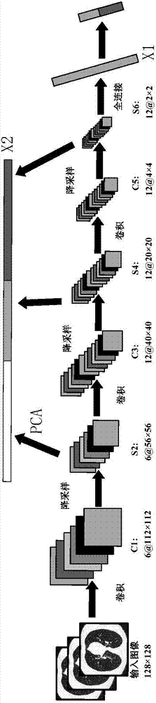
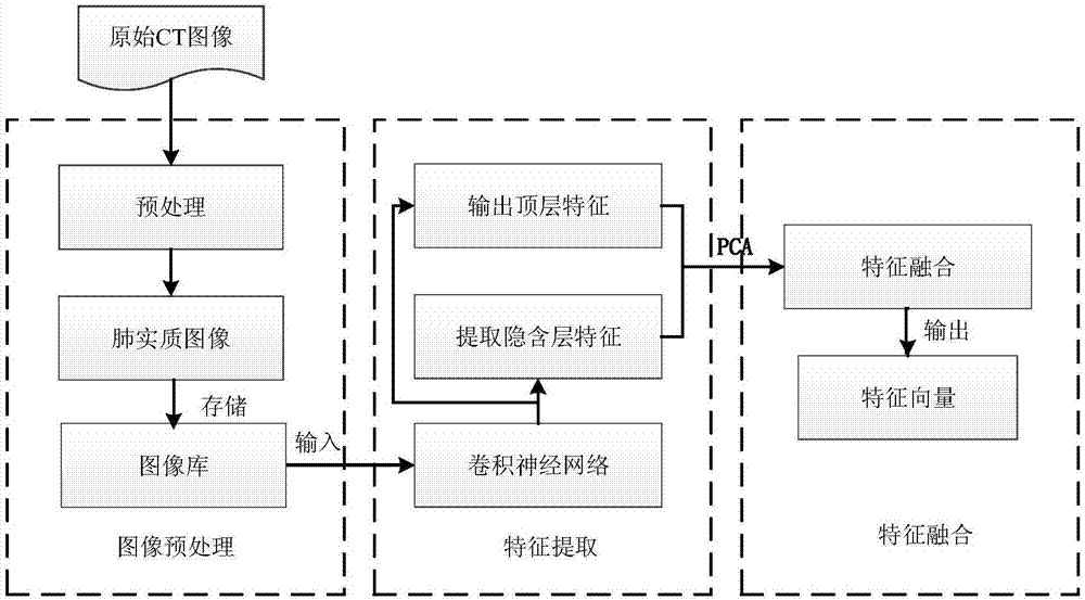
[0039] 1 Preprocessing of CT images

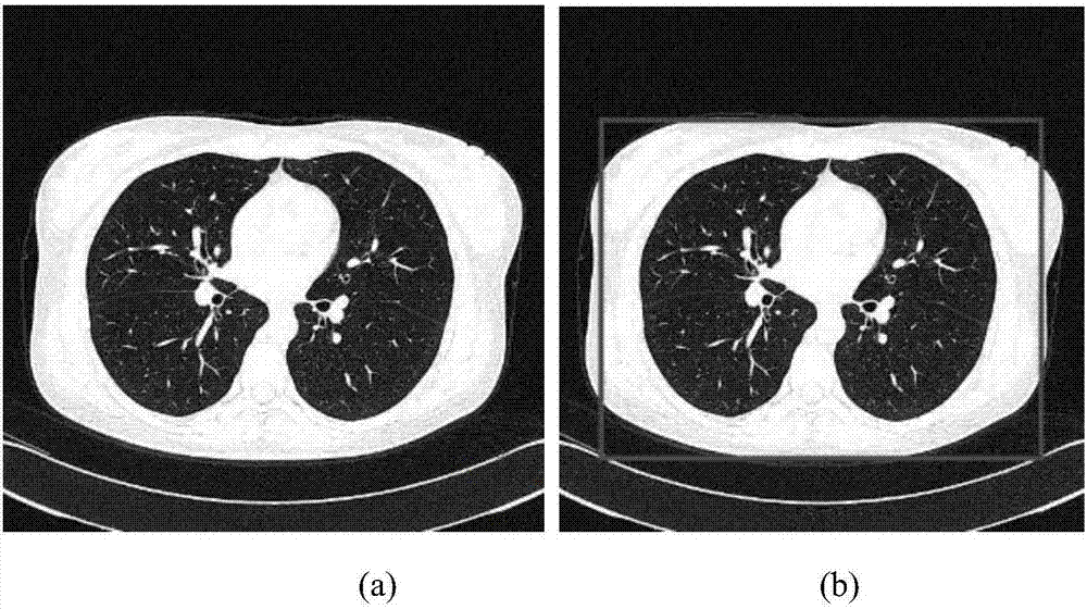
[0040] Before building the CNN model, the region of interest including the lung parenchyma was extracted from the CT image. The lung parenchyma is most complete when the lung CT sequence images take coordinates (40,110) in the upper left corner of the image and coordinates (470,440) in the lower right corner of the image. Such as figure 2 shown, yes figure 2 (a) Extract the lung parenchyma image to get figure 2 (b) Results shown. After the resulting image is normalized to a size of 112×112 through a two-line interpolation method, it is stored in the sample library for FeCNN training.

[0041] 2 feature extraction

[0042] When performing the feature extraction task of pulmonary nodules, FeCNN uses a group of units with the same weight vector but different positions on the thin-scan CT image to obtain the salient features of p...


PUM


Abstract

The invention discloses a pulmonary nodule feature extraction method based on a convolutional neural network and principle component analysis, which comprises the steps of firstly automatically learning pulmonary nodule features by using the convolutional neural network, performing feature extraction through convolution, and performing feature mapping through downsampling; and secondly, performing dimension reduction on the output of each feature mapping layer in a convolutional neural networks model for feature extraction (FeCNN) by using PCA (Principal Component Analysis) in feature extraction, and integrating with mapping of an output layer to acquire final multi-layer deep integration features. The pulmonary nodule feature extraction method not only can effectively recognize medical symptoms of a pulmonary nodule, but also avoids the complex feature extraction and feature reconstruction process in a traditional method, and plays a role of assisting diagnosis from an objective aspect.

Description

technical field [0001] The invention relates to a method for extracting features of pulmonary nodules based on a convolutional neural network and a principal component analysis method. Background technique [0002] The medical signs of pulmonary nodules are the basis for doctors to diagnose lung diseases. By analyzing various medical signs of lung CT images, it is convenient for doctors to judge the degree of benign and malignant nodules and make corresponding diagnostic decisions. However, doctors mainly diagnose diseases based on experience, and the diagnosis results are subject to some degree, so misdiagnosis and missed diagnosis often occur. In recent years, there is no need to manually extract features. Through the deep learning process, the data information hidden in the original input data can be extracted and abstracted layer by layer. The deeper the layer, the deeper the data concept represented by the extracted features. It cannot be expressed and obtained by the ...

Claims


Application Information

Patent Timeline
IPC IPC(8): G06T7/00G06K9/46
CPCG06T7/0012G06T2207/30064G06T2207/10081G06T2207/20084G06T2207/20081G06V10/44
Inventor 强彦肖小娇赵涓涓赵鹏飞王华
Owner TAIYUAN UNIV OF TECH
Why Eureka
Try Eureka