Automatic segmentation method for MRI image brain tumor based on full convolutional network

A fully convolutional network and automatic segmentation technology, applied in the field of medical image analysis, can solve problems such as low segmentation efficiency and rough segmentation results, and achieve the effects of improving efficiency, shortening training time, and saving data labeling costs

Active Publication Date: 2017-09-29
CHONGQING NORMAL UNIVERSITY
View PDF5 Cites 99 Cited by
  • Summary
  • Abstract
  • Description
  • Claims
  • Application Information

AI Technical Summary

Problems solved by technology

[0005] For the existing traditional segmentation method, it is necessary to understand the image through human subjective consciousness, so as to extract specific feature information, such as gray level information, texture information and symmetry information, to realize the segmentation of brain tumors. The result can only be better for specific images. Se

Method used


Image

  • Automatic segmentation method for MRI image brain tumor based on full convolutional network
  • Automatic segmentation method for MRI image brain tumor based on full convolutional network
  • Automatic segmentation method for MRI image brain tumor based on full convolutional network

Examples


Example Embodiment

[0044] In order to make it easy to understand the technical means, creation features, achieved goals and effects of the present invention, the present invention will be further described below with reference to the specific figures.

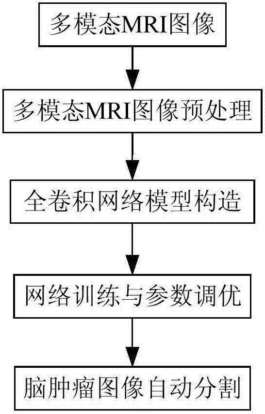
[0045] Please refer to figure 1 As shown, the present invention provides a fully convolutional network-based MRI image brain tumor automatic segmentation method, comprising the following steps:

[0046] S1. Brain tumor multimodal MRI image preprocessing, which includes:

[0047] S11. Perform a field offset correction operation on the two modal MRI images of T1 and T1c, specifically, the N4ITK method can be used to perform the offset field correction operation;

[0048] S12. Extract the MRI image slices of the four modalities of FLAIR, T1, T1c and T2. In each MRI image slice, set the highest gray value greater than 1% to 0.99 times the highest gray value, and set the highest gray value less than 1% to the lowest gray value. The intensity is set ...


PUM


Abstract

The invention provides an automatic segmentation method for an MRI (Magnetic Resonance Imaging) image brain tumor based on a full convolutional network. The method comprises multi-mode MRI image preprocessing of the brain tumor, construction of a full convolutional network model, network training and parameter optimization as well as automatic segmentation of a brain tumor image, specifically, the segmentation of the MRI image brain tumor is converted into a pixel-level semantic annotation problem and differential information emphasizing different modes of MRI, two-dimensional whole slices of four modes FLAIR, T1, T1c and T2 are synthesized into a four-channel input image, the convolutional layer and the pooling layer of the trained convolutional neural network are base feature layers, three convolutional layers equal to a full connection layer are added behind the base feature layers to form a middle layer, the middle layer outputs rough segmentation images corresponding to semantic segmentation types in quantity, and a de-convolutional network is added behind the middle layer and used for interpolating the rough segmentation images to obtain a fine segmentation image having the same size as the original image. The method does not need manual intervention, effectively improves the segmentation precision and efficiency, and shortens the training time.

Description

technical field [0001] The invention relates to the technical field of medical image analysis, in particular to a method for automatic segmentation of brain tumors in MRI images based on a fully convolutional network. Background technique [0002] Glioma is a common brain tumor that seriously threatens the lives of patients, and the most common treatment for brain tumors is surgical resection. MRI (Magnetic resonance imaging, MRI) displays the internal information of the brain in the form of images, and is a powerful tool for medical workers to analyze intracranial tumors. Brain tumor segmentation on MRI images plays a crucial role for early diagnosis, treatment planning, and treatment evaluation. However, the early manual segmentation and labeling methods were cumbersome and highly subjective. The boundary between glioma and normal tissue was not clear, and the MRI image itself was affected by noise, offset field effect, and partial volume effect. Therefore, designing an ...

Claims


Application Information

Patent Timeline
IPC IPC(8): G06T7/11G06T7/174G06N3/04G06N3/08
CPCG06N3/08G06T7/11G06T7/174G06T2207/10088G06T2207/30016G06T2207/30096G06N3/045
Inventor 崔少国毛雷熊舒羽刘畅
Owner CHONGQING NORMAL UNIVERSITY
Why Eureka
Try Eureka