Breast image processing method and breast imaging equipment

A breast and image technology, applied in the field of medical image processing, can solve problems such as uneven grayscale of breast images, and achieve the effect of reducing errors

Active Publication Date: 2021-04-23
SHANGHAI UNITED IMAGING HEALTHCARE
View PDF7 Cites 0 Cited by
  • Summary
  • Abstract
  • Description
  • Claims
  • Application Information

AI Technical Summary

Problems solved by technology

However, the above two methods are based on empirical thickness equalization, and the obtained breast images still have uneven gray levels, which has certain limitations.

Method used


Image

  • Breast image processing method and breast imaging equipment
  • Breast image processing method and breast imaging equipment
  • Breast image processing method and breast imaging equipment

Examples


Embodiment 1

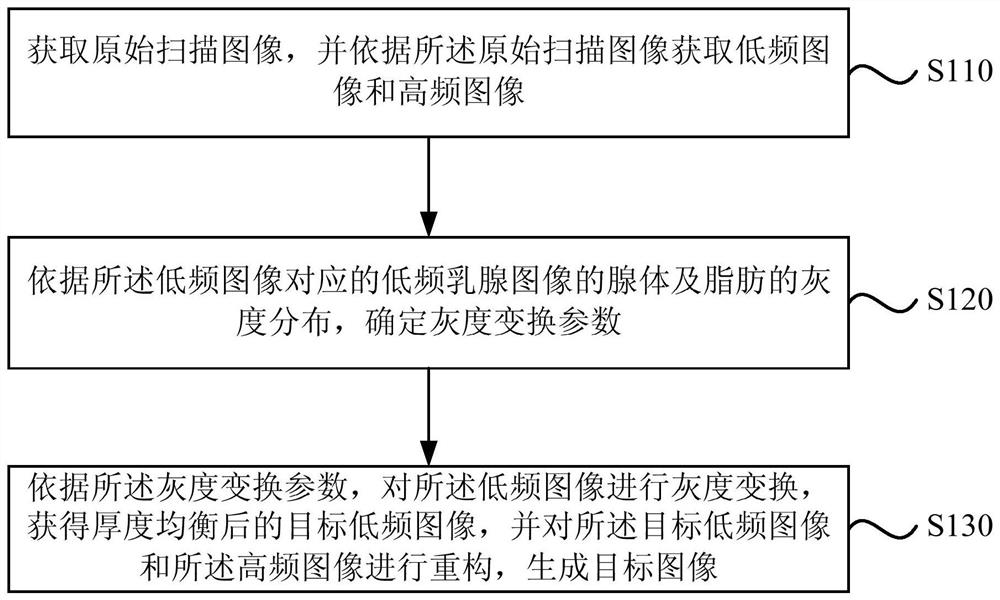
[0029] figure 1 It is a flowchart of a breast image processing method provided by Embodiment 1 of the present invention. The method can be performed by a breast image processing device, which can be realized by software and / or hardware, and which can be integrated in medical equipment capable of performing mammography, such as typically mammography equipment, such as mammography Dry plate X-ray photography system, dedicated screen film photography system or full-field digital mammography system (Full-Field Digital Mammography, FFDM), etc. Such as figure 1 As shown, the method of this embodiment specifically includes the following steps:

[0030] S110. Acquire an original scan image, and acquire a low-frequency image and a high-frequency image according to the original scan image.

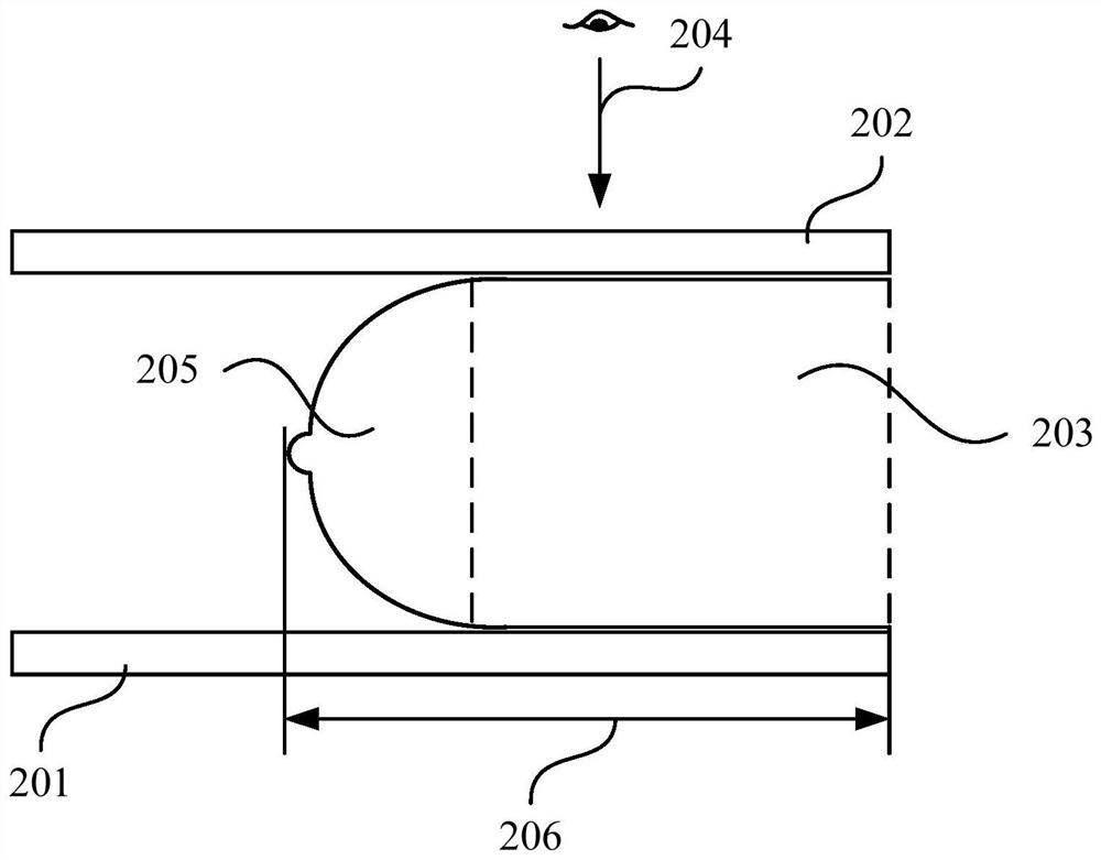
[0031] Specifically, such as figure 2 As shown, the mammary gland can be photographed using a mammography system such as FFDM. Firstly, use the support plate 201 and the compression plate 202 ...

Embodiment 2

[0049] Figure 7 It is a flowchart of a breast image processing method provided by Embodiment 2 of the present invention. On the basis of Embodiment 1 above, this embodiment further optimizes "determining grayscale transformation parameters according to the grayscale distribution". The explanations of terms that are the same as or corresponding to the above-mentioned embodiments will not be repeated here. The method of this embodiment includes:

[0050] S210. Acquire an original scan image, and acquire a low-frequency image and a high-frequency image according to the original scan image.

[0051] S220. Determine a gray scale transformation interval according to the maximum gray value of the breast image corresponding to the original scan image and the gray scale distribution of glands and fat in the low frequency breast image corresponding to the low frequency image.

[0052] Specifically, to determine the grayscale transformation parameters, a reasonable grayscale transform...

Embodiment 3

[0066] Figure 8 It is a flow chart of a mammary gland image processing method provided by Embodiment 3 of the present invention. On the basis of the above-mentioned Embodiment 2, this embodiment "according to the maximum gray value of the mammary gland image corresponding to the original scan image, the The low-frequency breast image corresponds to the gray-scale distribution of glands and fat in the low-frequency breast image, and the determination of the gray-scale transformation interval" has been further optimized. The explanations of terms that are the same as or corresponding to the above-mentioned embodiments will not be repeated here. The method of this embodiment includes:

[0067] S310. Acquire an original scan image, and acquire a low-frequency image and a high-frequency image according to the original scan image.

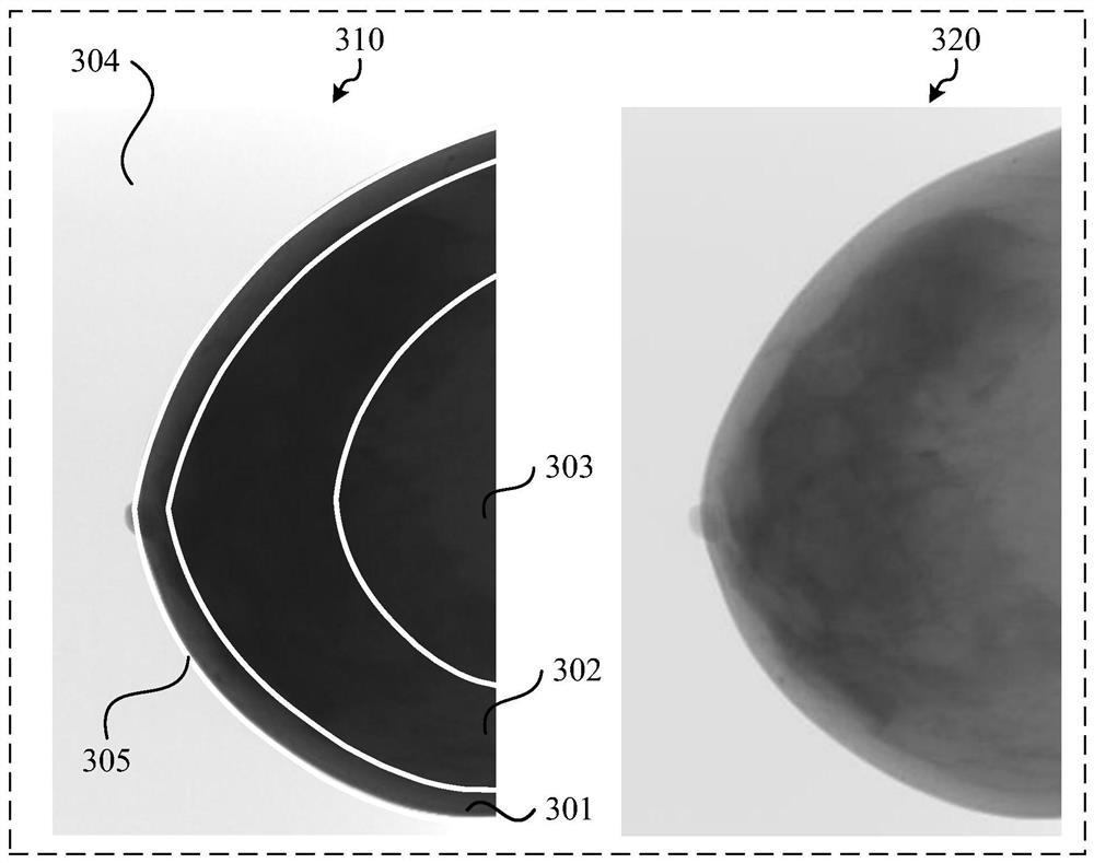
[0068] S320. Perform cropping processing on the low-frequency breast image corresponding to the low-frequency image to obtain a first low-frequency b...


PUM


Abstract

The embodiment of the invention discloses a mammary gland image processing method and a mammary gland imaging device. The method includes: obtaining an original scanning image, and obtaining a low-frequency image and a high-frequency image according to the original scanning image; determining gray-scale transformation parameters according to the gray-scale distribution of glands and fat of the low-frequency breast image corresponding to the low-frequency image; Perform grayscale transformation on the low-frequency image according to the grayscale transformation parameters to obtain a target low-frequency image after thickness equalization, and reconstruct the target low-frequency image and the high-frequency image to generate a target image. Through the above technical solution, the problem that breast image processing is too dependent on empirical parameters and its equalization effect is not good is solved, and the automation of breast image thickness equalization is realized to a certain extent, and breast images with more uniform gray scale distribution can be obtained, thereby Make breast images more in line with clinical needs.

Description

technical field [0001] Embodiments of the present invention relate to medical image processing technologies, and in particular to a breast image processing method and breast imaging equipment. Background technique [0002] During the imaging process of full digital mammography, the breast is photographed when the compression plate is compressed. However, since the compression plate cannot compress the edge of the mammary gland, the thickness of the mammary gland during shooting is uneven. This uneven thickness leads to a large difference in gray value between the fat at the edge and the fat at the inner side in the mammary gland image obtained by shooting. In this case, if you want to see all the breast tissue, the contrast of the whole breast image will be poor, which is not conducive to diagnosis; if you want to have a better contrast display, the fat at the edge may be outside the window width , cannot be displayed, resulting in loss of information, which is also not co...

Claims


Application Information

Patent Timeline
Patent Type & Authority Patents(China)
IPC IPC(8): G06T7/00G06T7/11G06T7/136G06T7/44G06T5/00
CPCG06T5/001G06T7/0012G06T2207/30068G06T7/11G06T7/136G06T7/44
Inventor 江春花赵书睿王汉禹
Owner SHANGHAI UNITED IMAGING HEALTHCARE
Why Eureka
Try Eureka