
A kit for detecting markers of ovarian cancer cells in peripheral blood

A technology of ovarian cancer cells and a kit, which is applied in the biological field and can solve the problems of being traumatic and unable to be used as an indicator for detecting tumor progress

Active Publication Date: 2021-04-02
牛刚 +1
View PDF2 Cites 0 Cited by
  • Summary
  • Abstract
  • Description
  • Claims
  • Application Information

AI Technical Summary

Problems solved by technology

The indicators of immune tissue detection include proliferating cell nuclear-associated antigen KI67, tumor suppressor gene p53, etc. The immunohistochemical results of ki67 become an indicator of tumor proliferation potential and an indicator of ovarian cancer malignancy, which can distinguish benign, borderline, and malignant ovaries In addition, its high expression in ovarian cancer is associated with poor prognosis, but it uses pathological tissue as a sample, which is relatively invasive and cannot be used as an indicator for detecting tumor progression. An effective detection method that can detect tumor progression in real time

Method used


Image

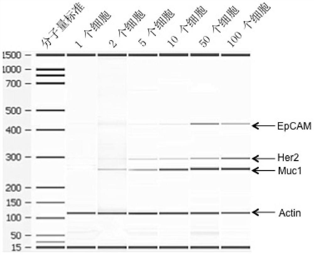
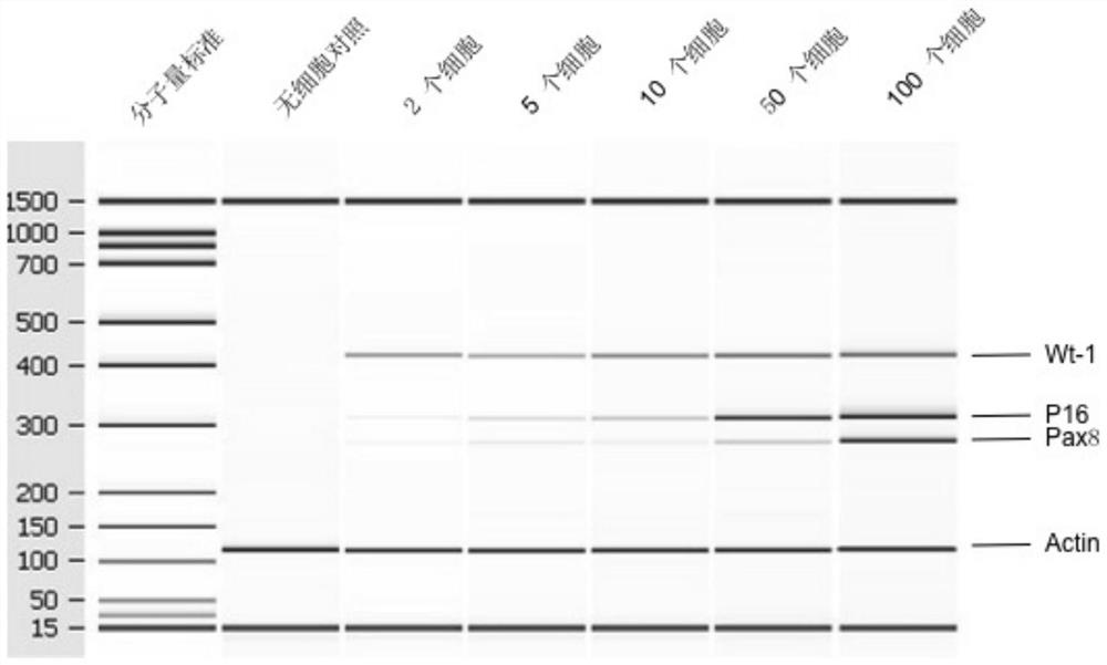
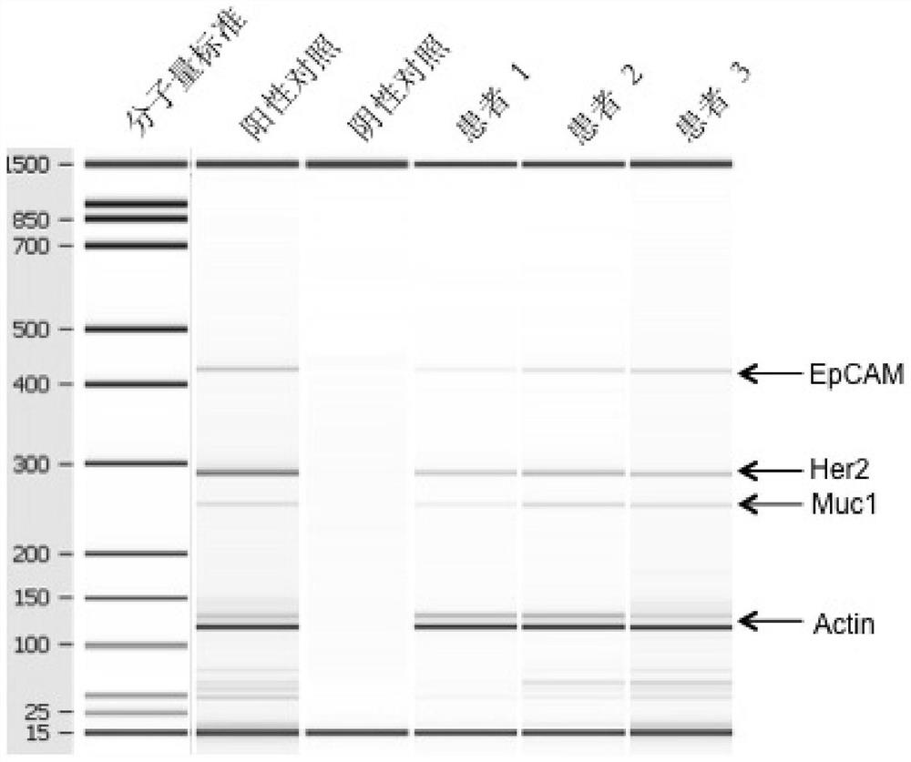
  • A kit for detecting markers of ovarian cancer cells in peripheral blood
  • A kit for detecting markers of ovarian cancer cells in peripheral blood
  • A kit for detecting markers of ovarian cancer cells in peripheral blood

Examples


Embodiment 1

[0080] Example 1: Preparation of magnetic beads bound to ovarian cancer antibody:

[0081] Antibodies used were anti-EpCAM monoclonal antibody, anti-Muc1 monoclonal antibody and anti-Her-2 monoclonal antibody. This kit uses the above three antibodies to be labeled separately, and then they are mixed for use.

[0082] Labeling method: magnetic beads produced by Invitrogen, USA M-450Tosylactivated. The marking method is strictly in accordance with the product instructions.

[0083] The labeling ratio is: label 500 μL magnetic beads per 100 μg antibody, and mix in equal amounts after labeling.

Embodiment 2

[0084] Example 2: Isolation of ovarian cancer cells:

[0085] 2.1 Sample handling:

[0086] Take 5mL of peripheral blood from patients with ovarian cancer, anticoagulate with EDTA, store at 4 degrees, and use within 48 hours.

[0087] 2.2 Magnetic bead treatment:

[0088] Wash the magnetic beads bound with ovarian cancer antibody (antibody-labeled magnetic beads, which are mixed magnetic beads labeled with three antibodies, and prepare antibody-labeled magnetic beads according to the ratio of 1 mL sample to 25 μL magnetic beads) with PBS repeatedly for three times, and separate the magnetic beads. Beads, kept on ice;

[0089] The specific operation is: draw 125μL of marked magnetic beads into a 1.5mL centrifuge tube, gently blow and mix with a pipette (note that a vortexer cannot be used), and then place it on the magnetic bead enricher for 1min. , to attach the magnetic beads to the wall of the centrifuge tube, discard the supernatant; then add 1mL PBS, place it on the mag...

Embodiment 3

[0096] Example 3: Detection of ovarian cancer cell markers:

[0097] 3.1 mRNA purification

[0098] Mix 40 μL magnetic beads with oligonucleotide Oligo(dT) into a 1.5 mL centrifuge tube, add 500 μL lysis / binding buffer to wash twice, separate magnetic beads, add supernatant, mix well, and incubate at room temperature 10min, let the mRNA bind to the magnetic beads; then separate the magnetic beads, use 500 μL buffer A to wash the magnetic beads twice, separate the magnetic beads, then wash the magnetic beads twice with 500 μL buffer B, separate the magnetic beads, and then use 100 μL RNase- Wash once with free water, separate the magnetic beads, resuspend the magnetic beads with 29.5 μL RNase-free water, incubate in a water bath (or metal bath) at 55°C for 5 min, and place on ice for 2 min to obtain the mRNA / magnetic bead mixture.

[0099] The mRNA / bead mix cannot be stored and needs to be reverse transcribed immediately.

[0100] 3.2 Reverse transcription:

[0101] The revers...


PUM


Abstract

The invention provides a kit for detecting markers of ovarian cancer cells in peripheral blood. The kit comprises magnetic beads and corresponding buffer reagents, wherein the magnetic beads are combined with ovarian cancer antibodies. According to the kit, the biological activity of tumor cells in blood circulation of a patient with ovarian cancer is detected through molecular biology methods including magnetic bead separation, RT-PCR and the like, and genes of a drug treatment target are determined; and by detecting the expression levels of EpCAM-2, Muc1, Her2, Wt-1, Pax8 and P16, the development and outcome of ovarian cancer are determined, and the theoretical basis and the experimental evidence are provided for the individual-based treatment of ovarian cancer. The kit provided by the invention is low in sample detection amount and high in detection sensitivity and has little harm to a patient, and the detection rate can reach up to 100%.

Description

technical field [0001] The invention belongs to the field of biotechnology, and relates to a kit for detecting ovarian cancer, in particular to a kit for detecting ovarian cancer cell markers in peripheral blood with high accuracy. Background technique [0002] 1.1 Overview of Ovarian Cancer [0003] Ovarian cancer (ovarian cancer) is one of the three malignant tumors of the female reproductive system. It is the most lethal gynecological tumor today. About 20,000 people are diagnosed with this disease every year, causing about 114,000 women worldwide to die of this disease every year. About 1 in 73 women will develop the disease in their lifetime. Epithelial ovarian cancer accounts for 90% of all ovarian tumors and can be divided into serous, mucinous, endometrioid, transparent There are five types of cell carcinoma (clear cell carcinoma) and transitional cell carcinoma (transitional cells carcinoma), among which serous ovarian cancer is the most, accounting for more than ...

Claims


Application Information

Patent Timeline
Patent Type & Authority Patents(China)
IPC IPC(8): C12Q1/6886C12Q1/6806C12N15/11G01N33/577G01N33/574
CPCC12Q1/6806C12Q1/6886C12Q2600/118C12Q2600/158G01N33/57449G01N33/577C12Q2563/143C12Q2563/149
Inventor 牛刚谭焕然
Owner 牛刚
Learn More