
Medical image aided diagnosis and semi-supervised sample generation system

A technology for assisting diagnosis and medical imaging, applied in neural learning methods, biological neural network models, special data processing applications, etc., it can solve the problems of inability to feedback closed-loop, self-learning, and inability to generate high-quality samples, so as to improve labeling efficiency , the effect of improving performance

Inactive Publication Date: 2018-01-09
杭州健培科技有限公司
View PDF2 Cites 39 Cited by
  • Summary
  • Abstract
  • Description
  • Claims
  • Application Information

AI Technical Summary

Problems solved by technology

This technology can be applied to a computer-aided diagnosis system based on deep learning to solve the problems that computer-aided software cannot feedback closed-loop, cannot generate high-quality samples, and cannot self-learn

Method used


Image

  • Medical image aided diagnosis and semi-supervised sample generation system
  • Medical image aided diagnosis and semi-supervised sample generation system
  • Medical image aided diagnosis and semi-supervised sample generation system

Examples


Embodiment Construction

[0033] The method of the present invention is further described below in conjunction with accompanying drawing and specific embodiment, and the specific steps of the present embodiment are as follows:

[0034] 1. Design steps

[0035] 1.1. Make training samples and test samples

[0036] 1.1.1. Obtain a large number of CT images: usually, the processed CT data layer thickness is 1.25-3mm, the layer spacing is 0.75-3mm, and the height (y) and width (x) of each layer of CT image are 512×512 pixels, The number of slices (z) of the CT image of a single case is 100-500, and the pixel size in the (z, y, x) direction is 0.5-1.5 mm;

[0037] 1.1.2. Adjust the appropriate HU window level: adjust the original CT image to an appropriate HU window level to eliminate the interference of other organs and tissues in the CT image. For example, when the area of ​​interest is lung tissue, the pixel with the HU value less than -1100 is usually assigned the value -1100, and the pixel with the HU...


PUM


Abstract

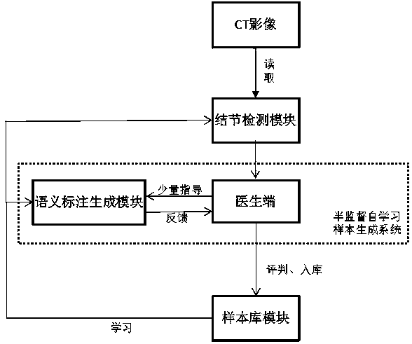
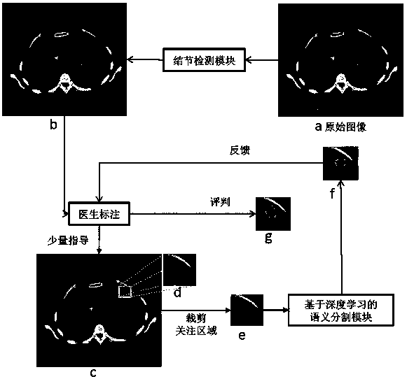
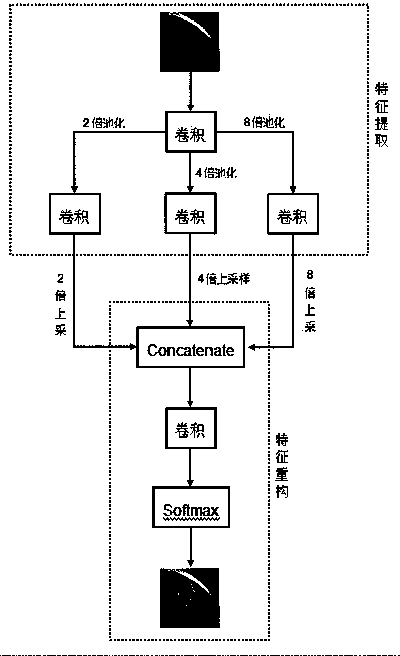
The invention relates to the technical field of medical image computer aided diagnosis, and discloses a medical image auxiliary diagnosis and semi-supervised sample generation system. The medical image aided diagnosis and semi-supervised sample generation system comprises a hospital image filing and communication module (a), a pulmonary nodule-based automatic detection module (b), a deep learning-based semantic annotation generation module (c) and a sample library module (d), wherein the hospital image filing and communication module is used for filing image data; the pulmonary nodule-based automatic detection module is used for automatically detecting pulmonary nodules to generate suspected pulmonary nodule positions; the deep learning-based semantic annotation generation module is used for receiving a small quantity of instructions of doctors and generating samples with accurate pulmonary nodule profiles and pulmonary nodule ingredient properties; and the sample library module is used for storing qualified samples so that online learning can be carried out by other self-learning systems. According to the system, the problem that computer aided software cannot feed closed loops back, cannot generate high-quality samples and cannot carry out self-learning is solved, and a feasible method is provided for a self-learning computer aided diagnosis system of CT images.

Description

technical field [0001] The invention relates to a system for medical image-aided diagnosis and semi-supervised sample generation, in particular to a technique for semi-supervisedly labeling precise semantic samples of pulmonary nodules using a small amount of guidance information from doctors. Background technique [0002] Electronic computerized tomography (ie CT scanning), using precisely collimated X-ray beams, gamma rays, ultrasonic waves, etc., together with highly sensitive detectors to scan a certain part of the human body one by one, has a fast scanning time. , clear images and other characteristics, the clinical results preliminarily prove that it is the most effective imaging method for detecting early asymptomatic lung cancer. In clinical diagnosis, a large number of lung diseases, including lung cancer, usually present as pulmonary nodules on CT images. Therefore, the detection and identification of pulmonary nodules using CT images is an important way to diagno...

Claims


Application Information

Patent Timeline
IPC IPC(8): G06K9/34G06K9/62G06N3/08G06F19/00
Inventor 程国华徐攀何林阳谢玮宜季红丽
Owner 杭州健培科技有限公司
Learn More