Heart image processing method in medical image

A medical image and image processing technology, applied in image data processing, image analysis, instruments, etc., can solve the problem of low accuracy of heart segmentation and achieve the effect of precise positioning

Active Publication Date: 2018-01-09
SHANGHAI UNITED IMAGING HEALTHCARE
View PDF7 Cites 14 Cited by
  • Summary
  • Abstract
  • Description
  • Claims
  • Application Information

AI Technical Summary

Problems solved by technology

[0004] Artifacts and noise caused by heart beats, and the proximity of the lungs, live

Method used


Image

  • Heart image processing method in medical image
  • Heart image processing method in medical image
  • Heart image processing method in medical image

Examples


Embodiment Construction

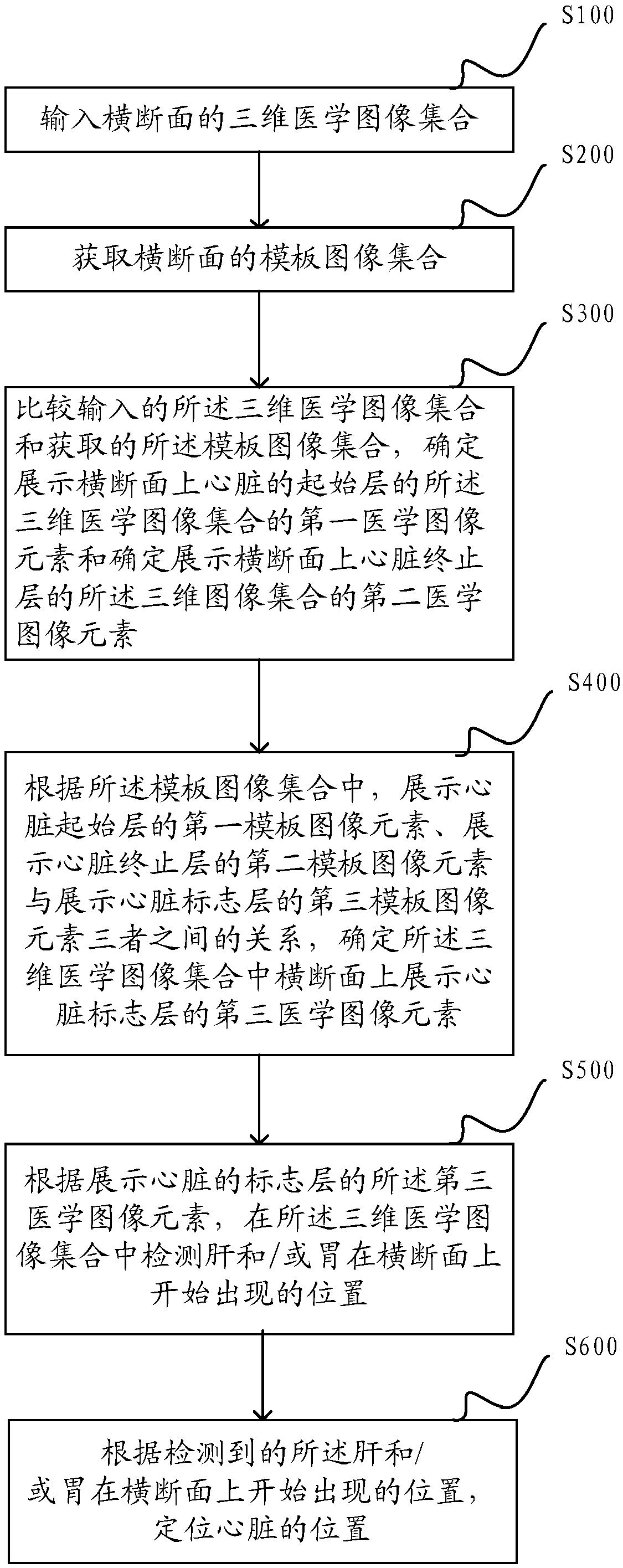
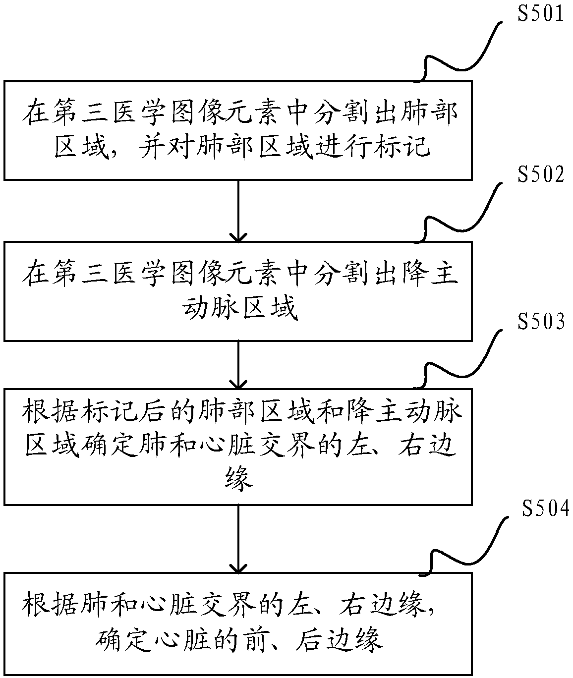
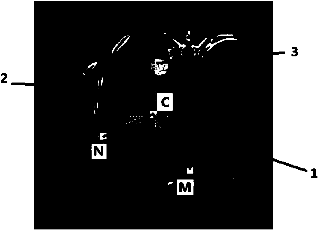
[0058] In order to make the purpose, technical solution and advantages of the present application clearer, the technical solution of the present application will be clearly and completely described below in conjunction with specific embodiments of the present application and corresponding drawings. Apparently, the described embodiments are only some of the embodiments of the present application, rather than all the embodiments. Based on the embodiments in this application, all other embodiments obtained by persons of ordinary skill in the art without making creative efforts belong to the scope of protection of this application.

[0059] In order to make the technology of the application easy to understand, the technology involved in the application is briefly described as follows:

[0060]Computed tomography (Computed Tomography, also known as computerized tomography, referred to as CT), is a diagnostic imaging examination. X-Ray Computed Tomography (X-CT for short) is a thre...


PUM


Abstract

The invention relates to a heart image processing method in a medical image. The method comprises the steps that the three-dimensional medical image set of a cross-section is input; the template imageset of the cross-section is acquired; the input three-dimensional medical image set is compared with the acquired template image set; a first medical image element, which demonstrates a heart start layer on the cross-section, of the three-dimensional medical image set and a second medical image element, which demonstrates a heart stop layer on the cross-section, of the three-dimensional image setare determined; and according to the relationship among a first template image element which demonstrates the heart start layer, a second template image element which demonstrates the heart stop layer, and a third template image element which demonstrates a heart marker layer in the template image set, a third medical image element which demonstrates the heart marker layer on the cross-section inthe three-dimensional medical image set is determined. According to the method provided by the invention, the heart marker layer can be accurately located, and the heart position can be accurately located.

Description

technical field [0001] The invention belongs to the technical field of image recognition and processing, in particular to a method for processing heart images in medical images. Background technique [0002] The heart is a key organ of the human cardiovascular system. Modern medical imaging techniques are able to provide structural and functional information of the heart. Among them, CT (Computed Tomography) has fast imaging speed, high resolution and large amount of information, and is an important means of heart examination. Segmentation of heart regions based on medical images, such as CT images, plays an important role in the diagnosis and treatment of cardiovascular diseases. At present, scholars at home and abroad have proposed many segmentation algorithms for certain anatomical structures in the heart, and these segmentation algorithms are mainly aimed at the segmentation of atrium and ventricle. For applications such as cardiac surgery navigation, interventional t...

Claims


Application Information

Patent Timeline
IPC IPC(8): G06T7/00G06T7/10
Inventor 姜娈李鹏程马金凤李强
Owner SHANGHAI UNITED IMAGING HEALTHCARE
Why Eureka
Try Eureka