A single domain antibody that recognizes hla-a2/rmfpnapyl

A technology for HLA-A2 and single-domain antibodies, which is applied in the field of single-domain antibodies that recognize HLA-A2/RMFPNAPYL, and can solve the problems of limited literature

Active Publication Date: 2020-07-07
SHENZHEN BEIKE BIOTECH
View PDF2 Cites 0 Cited by
  • Summary
  • Abstract
  • Description
  • Claims
  • Application Information

AI Technical Summary

Problems solved by technology

[0009] Most of the generated MAR molecules reported in the literature are essentially limited to specific recognition of MHC complexes at the cellular level or to be used to infer these complexes, with little literature demonstrating their practical application

Method used


Image

  • A single domain antibody that recognizes hla-a2/rmfpnapyl
  • A single domain antibody that recognizes hla-a2/rmfpnapyl
  • A single domain antibody that recognizes hla-a2/rmfpnapyl

Examples


Embodiment 1

[0051] Example 1 Screening the single domain antibody of HLA-A2 / RMFPNAPYL complex

[0052] 1.1 Single domain antibody phage library preparation

[0053] 1.1.1 Preparation of helper phage (BM13)

[0054] The M13KE phage replicon was double-digested with AlwnI and AfeI, and the artificially synthesized gene fragment was also double-digested with AlwnI and AfeI, and then ligated together with T4 ligase. After ligation, TG1 was transfected to obtain helper phage BM13. Thus, in the protoreplicon

[0055] The tctggtggtggttctggtggcggctctgagggtggtggctctgagggtggcggttctgagggtggcggctctgagggaggcggttccggtggtggctct sequence was replaced by a synthetic gene sequence, that is, a trypsin cleavage sequence was added in the phage GIII coding region. Once used as a helper phage, the trypsin digestion step was added to reduce the number of phages that did not contain the fusion target gene protein.

[0056] The synthetic gene sequence is as follows:

[0057] CCA GCC GGC CTT TCT GAG GGG TCG ACT...

Embodiment 2

[0102] Example 2, Expression of Single Domain Antibody

[0103] Prokaryotic single domain antibody expression

[0104] The four single domain antibody genes were cloned into pET22b with Nco I and Not I restriction endonucleases to express monovalent antibodies. The constructed vector was transformed into E.coli / DE3, single clones were picked the next day, and cultured with shaking at 220 rpm at 37°C until the OD600 was about 0.5. After adding IPTG (working concentration: 1 mM), the expression was induced at 220 rpm at 18°C ​​for 20 hours. Detection of protein expression. The sonication supernatant was purified with ProteinA and run SDS-PAG, see Figure 4 . Among them, the Marker strips are 14, 25, 30, 40, 50, 70, 100, 120, 160KD from small to large. Line1 is M5-H1, line2 is M5-G3, and line3 is M5-F4. Single domain antibodies are about 14KD in size.

[0105] Fusion FC expression in pET22b In order to form a bivalent antibody, the single domain antibody gene was connected ...

Embodiment 3

[0112] Example 3. Specific recognition of HLA-A2 / RMFPNAPYL complex by single domain antibody

[0113] Specific recognition of antigen complexes by single domain antibodies

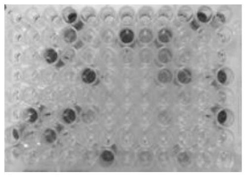
[0114] For the single-domain antibody expressed on pET22b, the cells were collected, resuspended evenly in PBS, and then sonicated. Ultrasonic crushing conditions: 600W, ultrasonic for 2 seconds, interval of 6 seconds, 10 minutes in total, 16°C. After ultrasonication, centrifuge at 12000rpm at 4°C for 10 minutes, take the supernatant for ELISA identification of the specificity of different antigens (Protein A-HRP is the secondary antibody) and read the plate. For data analysis, see Figure 9 . Wherein, the ordinate is the light absorption value at 650nm, and the abscissas 1, 2, 3, 4 are the four antigens HLA-A2 / ITDQVPFSV, HLA-A2 / NLVPMVATV, HLA-A2 / RMFPNAPYL, HLA-A2 / SLLMWITQC. The three samples are the ultrasonic supernatants of M5-H1, M5-G3, and M5-F4 expressed separately in pET22b.

[0115] The results...


PUM


Abstract

The invention discloses a single-domain antibody that recognizes HLA-A2 / RMFPNAPYL, and CDR1, CDR2 and CDR3 sequences for specific recognition; a fusion protein, which is characterized in that it uses the listed amino acid sequence and contains at least one other A fusion protein prepared from a polypeptide with a recognition function; a multi-specific or multifunctional molecule comprising the above-mentioned single-domain antibody; a nucleic acid molecule encoded as the above-mentioned polypeptide molecule; a carrier comprising the nucleic acid sequence; a The protein expressed by the nucleic acid molecule, its nucleotide sequence or at least a part of the sequence can be expressed through an appropriate expression system to obtain the corresponding protein or polypeptide; a host cell containing the nucleic acid sequence expressing the above protein. The invention not only recognizes the artificially synthesized HLA-A2 / RMFPNAPYL complex, but also can be combined with the HLA-A2 / RMFPNAPYL complex processed by natural antigens and expressed on the surface of tumor cells, which can be further developed into related tumor treatment products.

Description

technical field [0001] The invention belongs to the technical field of antibodies, in particular to a single-domain antibody for recognizing HLA-A2 / RMFPNAPYL and its application. Background technique [0002] The Wilms tumor gene (WT1) was discovered and cloned in Wilms tumor by different methods in 1990 by Call, Bonetta, Gessler and other three research groups. Highly expressed in tumors. Studies have shown that WT1 is a multifunctional transcriptional regulator that can regulate growth factors and receptors related to growth and differentiation, such as insulin-like growth factors (IGFs), insulin-like growth factor 1 receptor (IGF1R), platelet-derived growth factor A (PDGFA), epidermal growth factor (EGF), monocyte-macrophage colony-stimulating factor (M-CSF), multidrug resistance gene 1 (MDR1), etc. WT1 produces a variety of physiological effects by affecting its transcription, involving Cell growth, differentiation and apoptosis, etc. (ToskaE et al: Biochem J, 1:15, 20...

Claims


Application Information

Patent Timeline
Patent Type & Authority Patents(China)
IPC IPC(8): C07K16/46A61P35/00
CPCC07K16/18C07K16/2803C07K16/2833C07K2317/31C07K2317/565C07K2317/569
Inventor 古明珠高斌吕丽慧刘莹梁猛
Owner SHENZHEN BEIKE BIOTECH
Why Eureka
Try Eureka