
Diabetic retina image automatic classification method
An automatic classification and diabetes-based technology, which is applied in the direction of instruments, character and pattern recognition, and recognition of medical/anatomical patterns, etc., can solve problems such as poor classification performance, few retinal images, and difficult extraction of retinal image features to achieve accurate classification results , the effect of good robustness
- Summary
- Abstract
- Description
- Claims
- Application Information
AI Technical Summary
Problems solved by technology
Method used
Image

Examples
Embodiment Construction
[0019] See figure 1 , figure 2 , image 3 , a diabetic retinal image automatic classification method, the present invention is characterized in that:
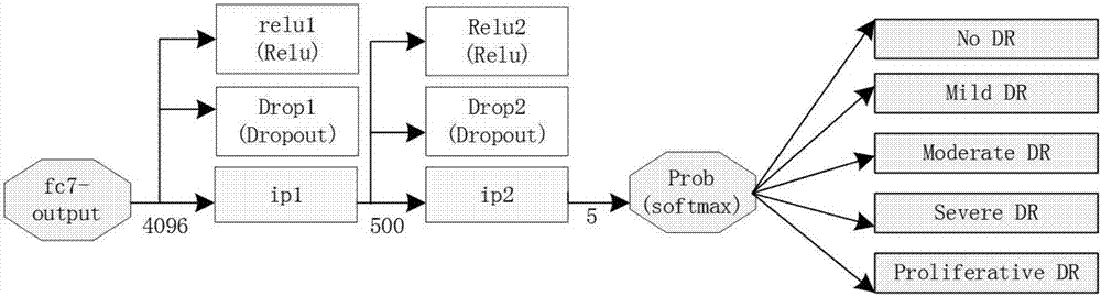
[0020] 1) The data comes from the Diabetic Retinopathy Detection competition in the data modeling and data analysis competition platform (kaggle). The retinal images in this data set are all high-resolution RGB images, and the retinal images are divided into normal and mild lesions according to the degree of lesions , moderate lesions, severe lesions, and proliferative lesions;
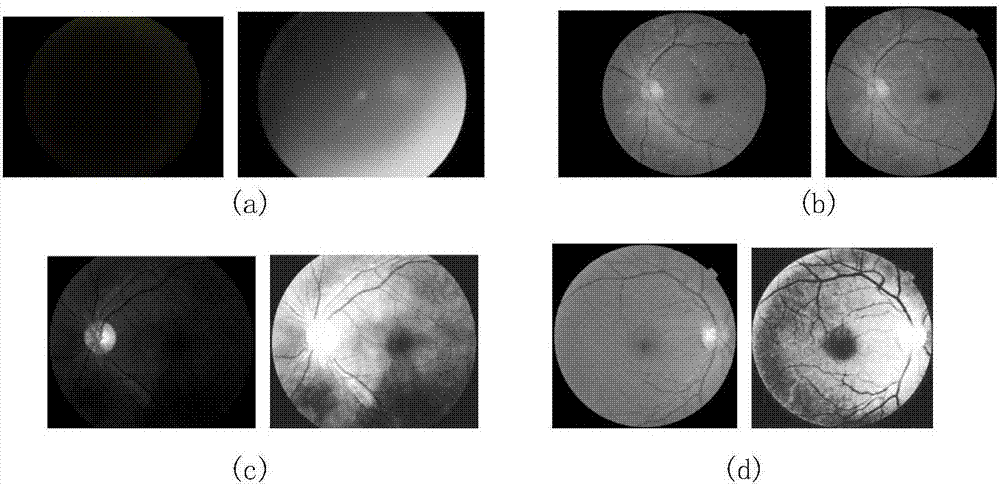
[0021] 2) Perform preprocessing such as denoising, histogram equalization, normalization, black border removal, data enhancement and feature analysis on the retinal image data;
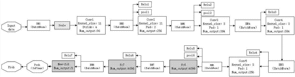
[0022] 3) In order to avoid problems such as slow convergence caused by changes in data distribution during the training process of the model, a BNnet is obtained by introducing a batch normalization layer before each convolutional layer and fully connected layer on the ba...
PUM
 Abstract
Description
Claims
Application Information