A MOS tube-based dual-gate regulated ultra-high sensitivity biosensor

A technology of biosensors and MOS tubes, which is applied in the direction of nanotechnology for sensing, instruments, scientific instruments, etc., can solve the limitations of the wide application of ultra-high sensitivity biosensors, uncontrollable width of silicon nanoribbons, unstable device performance, etc. To avoid the loss of tumor markers, meet the needs of subsequent detection applications, and reduce the cost of processing and use

Active Publication Date: 2019-04-23
WUXI PEOPLES HOSPITAL
View PDF6 Cites 0 Cited by
  • Summary
  • Abstract
  • Description
  • Claims
  • Application Information

AI Technical Summary

Problems solved by technology

[0004] However, the traditional wet etching process for preparing silicon nanoribbons, due to the uncontrollable etching rate, leads to unstable device performance, and the width of the obtained silicon nanoribbons is also uncontrollable, which eventually greatly increases the cost, which largely limits Extensive applications of ultra-high sensitivity biosensors

Method used


Image

  • A MOS tube-based dual-gate regulated ultra-high sensitivity biosensor
  • A MOS tube-based dual-gate regulated ultra-high sensitivity biosensor
  • A MOS tube-based dual-gate regulated ultra-high sensitivity biosensor

Examples


Embodiment 1

[0050] Embodiment 1: Preparation of sensor of the present invention

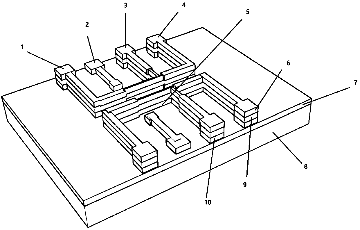
[0051] Such as figure 1 As shown, the MOS tube-based biosensor provided by the present invention, the detection system and the microfluidic channel system bonded to each other, the detection system includes a substrate 8 and an ion implantation layer 7 tiled above the substrate 8, the Two groups of U-shaped electrode groups facing away from each other are set on the ion implantation layer 7; the two wings of the U-shaped electrode group are source-drain electrodes 1, 4, and a top layer gate 3 is connected to the bottom of the U-shaped electrode group, and the A surface grid 2 parallel to the top grid 3 and not connected to the U-shaped electrode group is arranged inside the U-shaped electrode group;

[0052] The source-drain electrodes 1, 4 and the top gate 3 are sequentially composed of a silicon layer 10, an oxide layer 9 and a metal layer 6 upward from the ion implantation layer 7;

[0053] The bottom o...

Embodiment 2

[0076] Example 2: Trace and real-time detection of alpha-fetoprotein (AFP) and carcinoembryonic antigen (CEA):

[0077] Test according to the following methods:

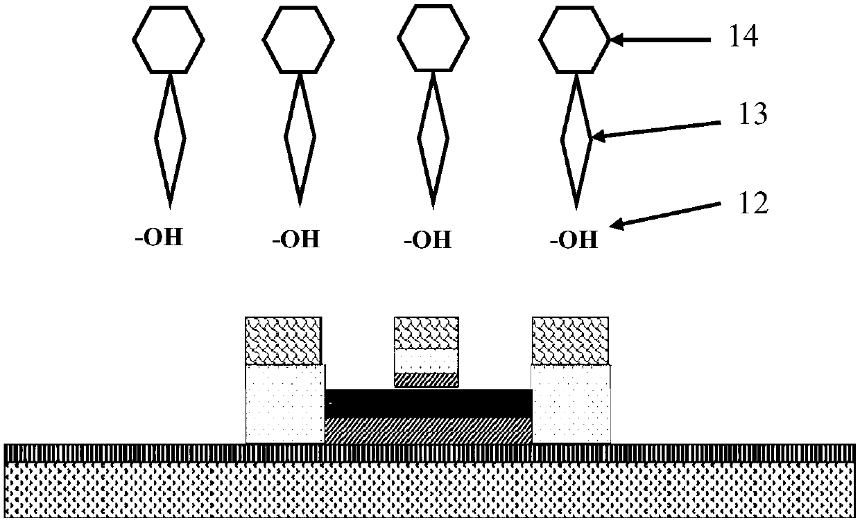
[0078] A. Modification of antibody protein: connect the microfluidic channel, use a syringe pump or a peristaltic pump to pass 1-1000 μg / ml antibody at room temperature and stay on the surface of the silicon nanowire 5, and the modification time is less than 0.1-10 hours; Washing with dyeing washing solution / PBST solution, drying the biosensor with nitrogen gas, the purpose of these operations is to modify the corresponding antibody of the target tumor marker on the silicon nanowire 5 of the biosensor;

[0079] B. Analysis: After fixing, pricking, and connecting pathways on the probe station, use a syringe pump or a peristaltic pump to pass the PBS solution through the microchannel system for 1-100 minutes at a flow rate of 0.001-100ml / min to obtain the basic current value. Then slowly transport the sample to be teste...


PUM

PropertyMeasurementUnit
thicknessaaaaaaaaaa
lengthaaaaaaaaaa
thicknessaaaaaaaaaa

Abstract

The invention discloses a MOS tube-based dual-gate control ultrahigh-sensitivity biosensor, which is applicable to the detection of a series of early tumors. The sensor is manufactured using SOI sheet processing, and a unique double-gate control structure is realized through ion implantation technology. The sensor is prepared by ultraviolet lithography combined with NLD etching method, which realizes the trace, instant and label-free detection of tumor markers. Compared with common sensors that detect current or conductance, this sensor detects changes in capacitance in the channel during antigen-antibody binding. The detection method involved in the present invention is more stable and has strong anti-interference ability, and can meet the requirements of detection range and sensitivity, especially in detection sensitivity. The minimum detectable concentration is in the range of 1fg / ml~1ng / ml samples within.

Description

technical field [0001] The invention relates to the technical field of biological material detection, in particular to a high-success-rate preparation and application method of an ultra-high-sensitivity biosensor based on a MOS tube. Background technique [0002] Malignant tumors are currently one of the major diseases that threaten human health, but most of the tumors are in the advanced stage of clinical manifestations, so early, rapid and sensitive diagnosis of them is an important way to improve the quality of human life. At present, the monitoring methods of clinical tumor incidence mainly rely on imaging examination and tumor marker detection. Imaging examinations often do not allow long-term follow-up due to resolution and radiation risks. Although the detection of tumor markers is relatively simple to operate, the sensitivity and specificity of tumor marker detection have become important factors that limit its application. Therefore, finding a simple and accurate ...

Claims


Application Information

Patent Timeline
Patent Type & Authority Patents(China)
IPC IPC(8): G01N33/574G01N33/543G01N27/22B82Y15/00B82Y40/00
CPCG01N27/227G01N33/54366G01N33/57484B82Y15/00B82Y40/00G01N33/5438G01N33/574G01N27/4146G01N27/4145G01N33/57473G01N33/57476H01L21/02057H01L21/02164H01L21/0217H01L21/022H01L21/02238H01L21/02274H01L21/02532H01L21/02603H01L21/0274H01L21/26513H01L21/266H01L21/28079H01L21/28194H01L21/28568H01L21/3065H01L21/308G01N33/54373
Inventor 王彤
Owner WUXI PEOPLES HOSPITAL
Why Eureka
Try Eureka