Synchronous display system for sectional images and three-dimensional anatomic images of medical images

A technology of tomographic images and medical imaging, which is applied in the field of medical display, can solve the problems of understanding relevant tomographic anatomy, inaccurate grasp of image anatomical structure, learning, etc.

Inactive Publication Date: 2018-09-18
徐州云联医疗科技有限公司
View PDF9 Cites 5 Cited by
  • Summary
  • Abstract
  • Description
  • Claims
  • Application Information

AI Technical Summary

Problems solved by technology

[0003] Most of the existing technical means revolve around the matching of standard images and test images, and use VR technology to realize three-dimensional virtual image display; and in medical anatomy teaching, the two-dimensional tomographic image of the image is not synchronized with the 3D image of the general anatomy of the human body, and Real-time display is realized, and it is impossible to realize the transformation and rotation of any angle in the space, and the relationship with the surrounding adjacent structures, so that medical students and junior doctors cannot learn and understand the relevant common sense of tomographic anatomy vividly and vividly, and cannot There is a problem that low-level imaging diagnosticians and clinicians are not sure about the anatomical structure in the image when interpreting the tomographic image

Method used


Image

  • Synchronous display system for sectional images and three-dimensional anatomic images of medical images
  • Synchronous display system for sectional images and three-dimensional anatomic images of medical images
  • Synchronous display system for sectional images and three-dimensional anatomic images of medical images

Examples


Embodiment 1

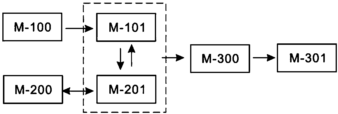
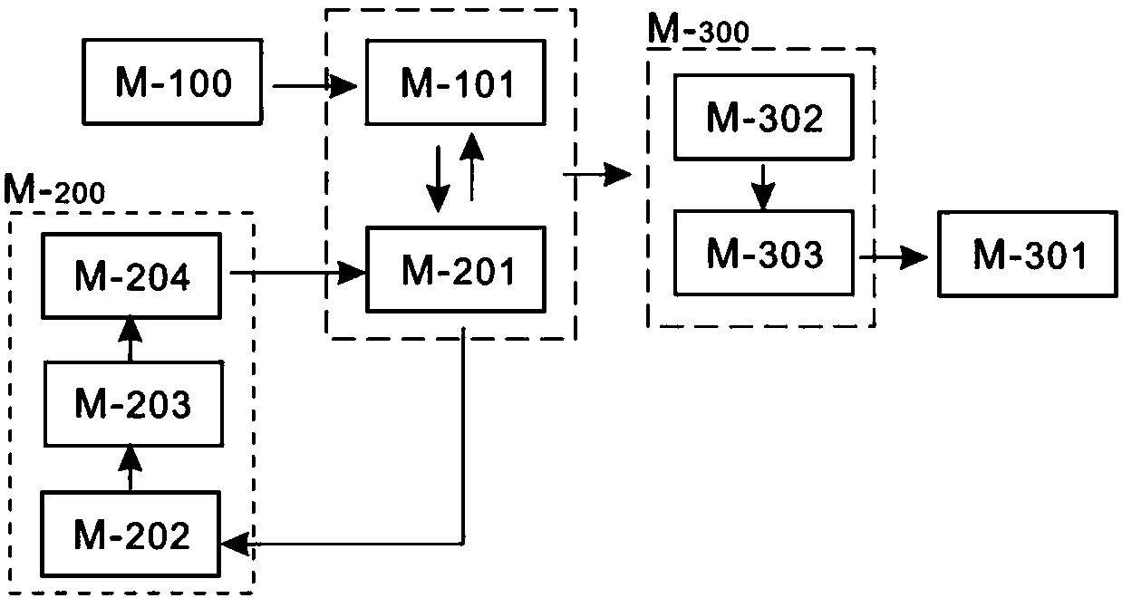
[0041] refer to Figure 1~2 In order to realize the synchronous real-time display of the two-dimensional tomographic image and the 3D image of the general anatomy of the human body, and provide the anatomical name of the target tissue or organ, in this embodiment, the synchronous display system of the tomographic image of the medical image and the three-dimensional anatomical image includes An image acquisition device M-100, an image matching module M-200 and a display module M-300. Specifically, the image acquisition device M-100 acquires the continuous standard axial images M-101 from the head to the feet by scanning the normal human body, and sets the corresponding window width and window level at the corresponding parts; in this implementation In the example, the image acquisition device M-100 includes CT or MR, and scans the normal human body under the conditions of 120KV, 80mAs, slice thickness 1mm and slice distance 1mm to obtain the standard axial image M-101, where CT...

Embodiment 2

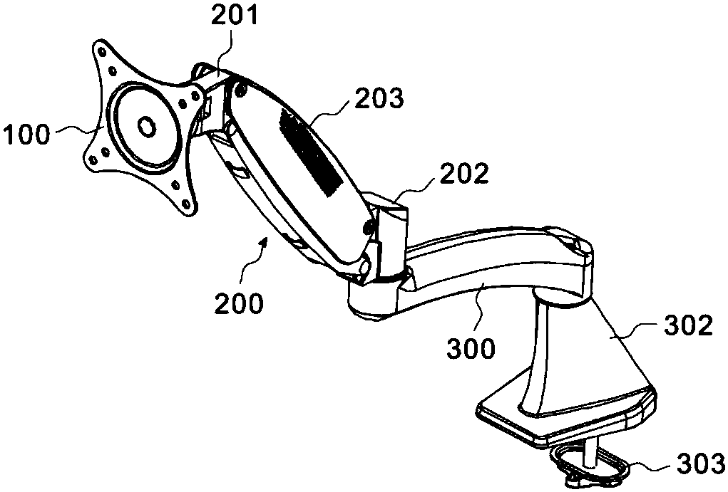
[0046] This embodiment provides an application display device that realizes the above-mentioned display interface M-301, such as a display screen or display A, and the display A is used in the synchronous display system, in order to facilitate the operator to view the content of the display A from various angles , obtain the signal transmitted by the display A in time, and make corresponding countermeasures. In this embodiment, a display stand is also provided. The existing display A is usually fixed to the wall, the ground, the desktop or other frames through the display stand. . A general display bracket usually includes a mounting panel connected to the back of the display device and a bracket fixed to the mounting panel. The bracket includes a wrist joint fixed to the installation panel, an arm joint movably connected with the wrist joint, etc.; the wrist joint and the arm joint are manually rotated to adjust the angle of the display A to facilitate people's viewing. In th...

Embodiment 3

[0052]In the above-mentioned first embodiment, relative rotation can occur between the shaft seat 103 and the shaft pin 104, and the horizontal shaft block 201a is inserted into the shaft pin 104 to limit and fix, and there will be no relative rotation between the two, so the installation The disk 100 can rotate around the horizontal axis block 201a, but if the weight of the display screen on the installation disk 100 is too large, and with the increase of the number of rotations between the shaft pin 104 and the shaft seat 103, the contact surface will gradually change after continuous wear. It is smooth and the friction force gradually decreases, so the mounting plate 100 cannot be fixed at the designated position during use, and a slight vibration will cause the mounting plate 100 to slide down, so the difference between this embodiment and the first embodiment That is: a magnetic lock module 400 is also provided between the shaft pin 104 and the shaft seat 103, which can lo...


PUM


Abstract

The invention discloses a synchronous display system for sectional images and three-dimensional anatomic images of medical images. The device includes an image acquisition device, an image matching module and a display module which can synchronously display standard axial position images and three-dimensional general anatomic structure images of a human body on a display interface in real time anddisplay names matching corresponding target tissues and organs. The beneficial effects of the synchronous display system are that two-dimensional sectional images and the human body gross anatomy 3Dimages of the images can be synchronously displayed in real time, 3D spatial whole anatomy of the organs / tissues can be displayed at the same time, the transformation and rotation of arbitrary anglesin space and relationship with adjacent structures can be realized, and the visualization of the two-dimensional image sectional images to the three-dimensional stereo spatial images can be realized.

Description

technical field [0001] The invention relates to the technical field of medical display, in particular to a synchronous display system of a medical imaging tomographic image and a three-dimensional anatomical image. Background technique [0002] With the continuous development of science and technology in recent years, rapid progress has been made in the field of medical research at this stage. Medicine is a subject that deals with various diseases or lesions of the human body through scientific or technical means; it is an applied subject of biology, divided into basic medicine and clinical medicine; it is an advanced science that deals with human diseases from the anatomical and molecular genetic levels; it It is a systematic discipline from prevention to treatment of diseases, and its research fields include basic medicine, clinical medicine, forensic medicine, laboratory medicine, preventive medicine, health care medicine, rehabilitation medicine, etc. Human anatomy is a...

Claims


Application Information

Patent Timeline
Patent Type & Authority Applications(China)
IPC IPC(8): A61B5/00
CPCA61B5/7425
Inventor 李晓峰胡延军李曾
Owner 徐州云联医疗科技有限公司
Why Eureka
Try Eureka