Method for establishing mouse model of cardiac volume overload

A model and capacity technology, applied in the field of biomedicine, can solve problems such as reflux, high mortality, and massive bleeding, and achieve convenient operation and good repeatability

Inactive Publication Date: 2018-09-21
ZHONGSHAN HOSPITAL FUDAN UNIV
View PDF0 Cites 1 Cited by
  • Summary
  • Abstract
  • Description
  • Claims
  • Application Information

AI Technical Summary

Problems solved by technology

However, because the position of the aortic valve is not accurately known, the pressure catheter often punctures the aorta and causes massive bleeding; and the tip of the pressure catheter is relatively thick compared to the mouse aortic valve, which can easily cause extremely severe regurgitation , resulting in a significant increase in the mortality rate
Because of the high mortality rate and low success rate, by searching Pubmed and Google Scholar, it can be found that these two research groups have not reported the mouse aortic regurgitation model since then

Method used


Image

  • Method for establishing mouse model of cardiac volume overload
  • Method for establishing mouse model of cardiac volume overload
  • Method for establishing mouse model of cardiac volume overload

Examples


Embodiment 1

[0055] Example 1 Establishment of cardiac volume overload mouse model

[0056] For mice undergoing aortic valve regurgitation surgery (true surgery), after intraperitoneal anesthesia with ketamine (150 mg / kg) and xylazine (10 mg / kg), the mice were fixed on a heating plate, and the ends of the limbs of the mice were smeared with The conductive paste is then attached to the conductive area of ​​the heating plate. Subsequently, a 27G cannula 2 with a built-in 0.16mm diameter steel wire 1 was inserted through the right common carotid artery (RCCA) ( figure 1 with 2 shown). Under the guidance of high-frequency ultrasound, the root of the aortic valve is clearly displayed, the cannula is advanced to the front of the aortic valve, and the built-in steel wire is stretched out from the cannula to puncture part of the aortic valve ( figure 2 shown). During the modeling process, the Doppler ultrasound of the blood flow at the aortic arch was monitored at any time, and the modeling w...

Embodiment 2

[0057] Embodiment 2 Realization of Quantitative Control of Reflux Degree

[0058] Non-invasive and real-time monitoring of aortic arch blood flow by high-frequency ultrasound of small animals can quantify the degree of reflux, and according to the needs of experiments, mouse models with different degrees of reflux can be produced ( Figure 4 ). This is a huge advantage over previous arteriovenous fistula volume overload models that cannot quantify the severity of stoma after stoma.

Embodiment 3

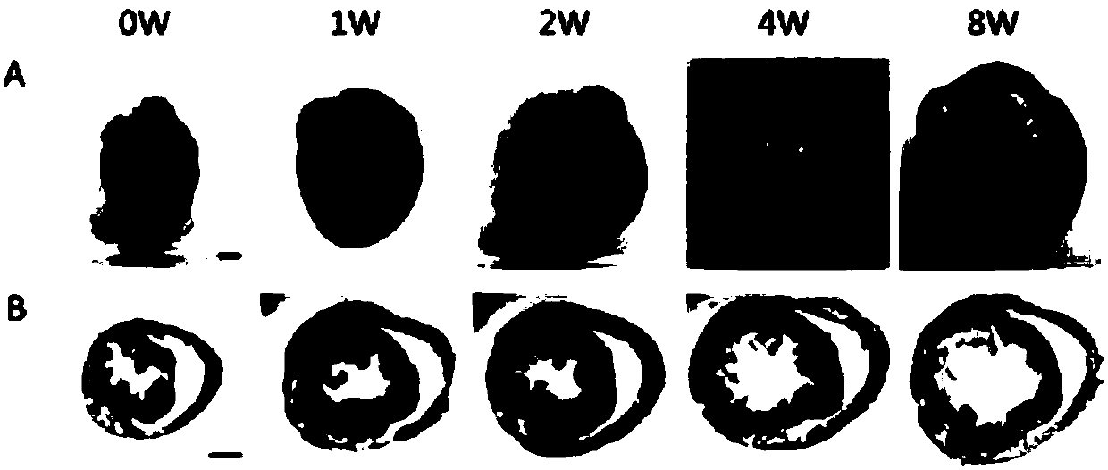
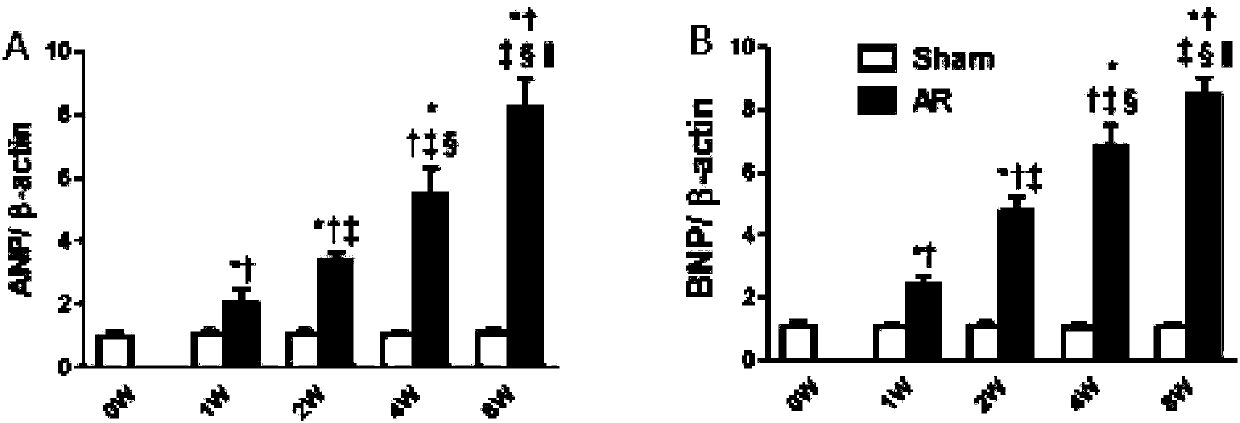
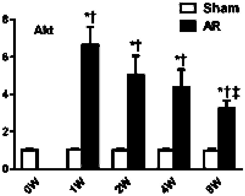
[0059] Example 3 Changes in Left Heart Structure and Cardiac Function of Mice Before and After 1 Week, 2 Weeks, 4 Weeks and 8 Weeks in Moderate Reflux State

[0060] Figure 5 It is shown that the reverse blood flow of the aortic arch of mice at 1 week, 2 weeks, 4 weeks, and 8 weeks after operation is obvious, and there is no obvious change at each time point after operation; Image 6 Shown are the changes of the long-axis M-mode ultrasonography of the mouse left ventricle before operation and 1 week, 2 weeks, 4 weeks, and 8 weeks after operation. It can be seen that the left ventricular diameter is significantly enlarged; Figure 7 Significant increase in left ventricular end-diastolic diameter, Figure 8 Significant increase in left ventricular end-systolic diameter; Figure 9 A slight increase in left ventricular end-diastolic posterior wall thickness two weeks after surgery, Figure 10 The left ventricular end-systolic posterior wall thickness increased slightly two wee...


PUM


Abstract

The invention discloses a method for establishing a mouse model of cardiac volume overload. Under guidance of high-frequency ultrasounds, metal wires of a built-in metal wire cannula used for a right-side common carotid artery damage an aortic valve once or multiple times, thereby causing blood regurgitation of the aortic valve. During a model establishing process, doppler ultrasounds of blood regurgitations at arcus aortae are monitored any time till the regurgitation degree is satisfied. The method enjoys obvious advantages over a present model-establishing method for arterio-venous fistulas. The method can establish a mouse model for aortic regurgitation with convenient operation, good regurgitation and repetitiveness and provide a novel and model animal satisfactory to simulated clinical lesions for the study of translational medicine for cardiac volume overload reconstitution. The method and a model of pressure overload can also be used for differential expressions of cardiac remodeling, which lay a foundation for clarification of a molecular mechanism for two types of overload cardiac remodeling differences. The method can provide importance references for personalized diagnosis and treatment of different types of clinical cardiomegaly diseases.

Description

technical field [0001] The invention belongs to the technical field of biomedicine, and in particular relates to a method for establishing a cardiac capacity overload model and evaluating it by destroying the mouse aortic valve under the guidance of high-frequency ultrasound. Background technique [0002] Cardiovascular disease has gradually become the leading cause of morbidity and mortality in western developed countries and in my country, causing serious social and economic burdens. It is particularly urgent and necessary to discover the pathogenesis of cardiovascular disease from the source and carry out targeted prevention and treatment accordingly. The mechanical overload of the heart is manifested by the heart being subjected to abnormally elevated mechanical stress, which is the cause or complication of various cardiovascular diseases, which directly triggers cardiac remodeling. Mechanical stress overload can be divided into two categories: pressure overload and vol...

Claims


Application Information

Patent Timeline
Patent Type & Authority Applications(China)
IPC IPC(8): A61D1/00A61B8/06
CPCA61B8/06A61B8/488A61B34/20A61B2034/2063A61D1/00
Inventor 吴剑邹云增游洁芸王时俊黄家园丁志文杨春杰康乐姜红
Owner ZHONGSHAN HOSPITAL FUDAN UNIV
Why Eureka
Try Eureka