
Systems and methods for removing occluded objects in surgical images and/or videos

An image removal technology, applied in surgery, image enhancement, image analysis, etc., can solve problems such as damage and blurred lenses

Active Publication Date: 2021-06-01
COVIDIEN LP
View PDF5 Cites 0 Cited by
  • Summary
  • Abstract
  • Description
  • Claims
  • Application Information

AI Technical Summary

Problems solved by technology

In the case of three-dimensional (3D) endoscopy, when the tool is too close to the lens and / or the tool is moving, the user may experience disorientation and / or nausea, thereby forcing the user to limit their movement options
Additionally, localized contamination may blur the lens, thereby compromising portions of any image obtained by the endoscope

Method used


Image

  • Systems and methods for removing occluded objects in surgical images and/or videos
  • Systems and methods for removing occluded objects in surgical images and/or videos
  • Systems and methods for removing occluded objects in surgical images and/or videos

Examples


Embodiment Construction

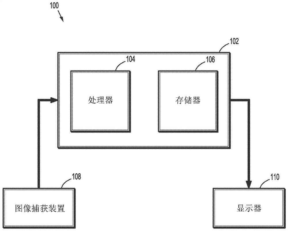
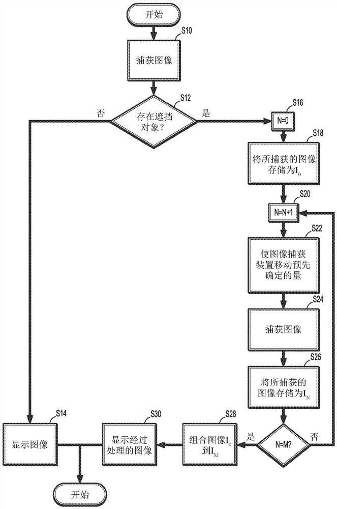
[0029] Image data captured from the endoscope during a surgical procedure can be analyzed to remove occluding objects from the endoscope's viewpoint. Various images of the surgical site can be obtained by moving the endoscope according to a predetermined scheme. The various images can then be combined to present a single image or video to the user with the occluding object removed from the image or video.

[0030] One or more of these techniques may be incorporated as part of the imaging system in the surgical robotic system to provide the clinician with additional information within the field of view of the endoscope. This enables clinicians to quickly identify, avoid and / or correct inappropriate situations and conditions during surgery.

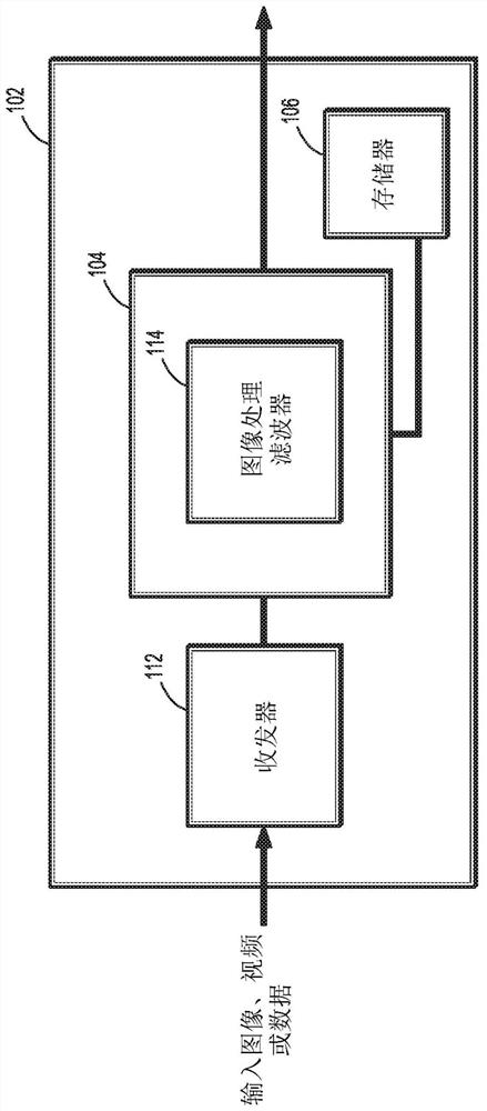
[0031] The present disclosure relates to systems and methods for providing processed images in real-time to a clinician during a surgical procedure with occluding objects removed from the images. The systems and methods described herein a...


PUM


Abstract

The present disclosure relates to systems and methods for removing occluding objects from surgical images. An image capture device is inserted into a patient and captures initial images of a surgical site within the patient during a surgical procedure. A controller receives the image and determines that the occluding object exists in the initial image. The controller executes a removal algorithm comprising controlling the image capture device to perform a plurality of movements, controlling the image capture device to capture a plurality of images, wherein each image in the plurality of images corresponding to a movement of the plurality of movements, and applying an image filter to combine the initial image and the plurality of images and generate a processed image, wherein the occluding object is moved from the processed image remove.

Description

[0001] Cross References to Related Applications [0002] This application claims the benefit and priority of U.S. Provisional Patent Application No. 62 / 302,366, filed March 2, 2016, the entire disclosure of which is incorporated herein by reference. Background technique [0003] Minimally invasive surgery involves performing a surgical procedure using multiple small incisions rather than one larger opening or incision. Small incisions reduce patient discomfort and shorten recovery time. Small incisions also limit visibility of internal organs, tissues, and other matter. [0004] Endoscopes have been used and inserted into one or more incisions to make it easier for a clinician to see internal organs, tissues and other substances in the body during surgery. These endoscopes contain a camera with optical and / or digital zoom capabilities coupled to a display that shows a magnified view of internal organs, tissues and substances captured by the camera. One or more surgical tool...

Claims


Application Information

Patent Timeline
Patent Type & Authority Patents(China)
IPC IPC(8): A61B90/00
CPCA61B34/76A61B90/00A61B90/361G06T5/50A61B34/30A61B34/37G06T2207/20212G06T2207/10068A61B2090/3612G06T5/77G06T2207/20221
Inventor 德怀特·梅格兰
Owner COVIDIEN LP
Learn More