
Ultrasound contrast imaging method and ultrasound imaging system

An ultrasonic imaging system and imaging method technology, applied in ultrasonic/sonic/infrasonic diagnosis, structure of ultrasonic/sonic/infrasonic diagnostic equipment, radio wave measurement system, etc. Unable to determine the contrast agent microbubble speed, low frame rate and other issues

Active Publication Date: 2018-11-23
SHENZHEN MINDRAY BIO MEDICAL ELECTRONICS CO LTD +1
View PDF5 Cites 8 Cited by
  • Summary
  • Abstract
  • Description
  • Claims
  • Application Information

AI Technical Summary

Problems solved by technology

In addition to the disadvantage of low frame rate, the existing line-by-line scanning contrast imaging technology can only show the difference in microbubble intensity on the image, and the speed difference of contrast agent microbubbles in the cut plane cannot be displayed intuitively. It is impossible to accurately determine the velocity of contrast agent microbubbles through ultrasound-contrast images

Method used


Image

  • Ultrasound contrast imaging method and ultrasound imaging system
  • Ultrasound contrast imaging method and ultrasound imaging system
  • Ultrasound contrast imaging method and ultrasound imaging system

Examples


Embodiment Construction

[0038] The following will clearly and completely describe the technical solutions in the embodiments of the present invention with reference to the accompanying drawings in the embodiments of the present invention. Obviously, the described embodiments are only some of the embodiments of the present invention, not all of them. Based on the embodiments of the present invention, all other embodiments obtained by persons of ordinary skill in the art without making creative efforts belong to the protection scope of the present invention.

[0039] Figure 5 It is a schematic structural block diagram of an ultrasound imaging system in an embodiment of the present invention. refer to Figure 5 , in some embodiments of the present invention, the ultrasound imaging system 100 may include a transducer 10 , a transmit / receive selection switch 80 , a transmit circuit 20 , a receive circuit 30 , a processor 40 , a display unit 60 , an input device 50 and a memory 70 . Transmit circuit 20 ...


PUM


Abstract

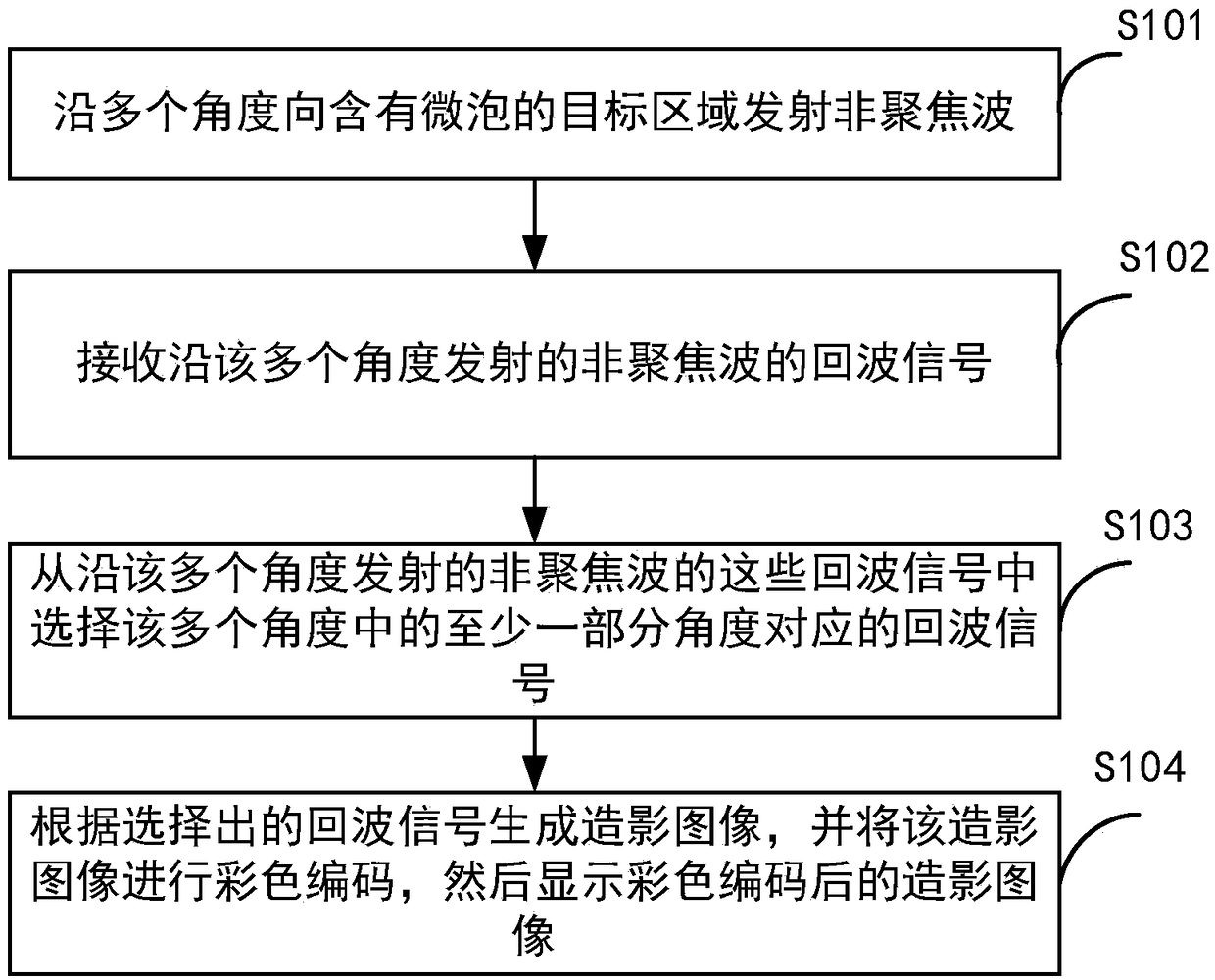
The present application discloses an ultrasound contrast imaging method comprising: transmitting an unfocused wave to a target region containing microbubbles in a plurality of directions; receiving echo signals of the unfocused wave; and selecting an echo signal corresponding to at least a portion of the angles in a plurality of angles from the echo signals; and generating a contrastographic picture based on the selected echo signal and displaying the same in color codes. The application also discloses an ultrasound imaging system.

Description

technical field [0001] The present invention relates to an ultrasound imaging method, in particular to an ultrasound contrast imaging method and an ultrasound imaging system applying the ultrasound contrast imaging method. Background technique [0002] Ultrasound contrast agent microbubbles can enhance the intensity of reflected echoes. Its diameter is very small, about the size of blood cells, and can diffuse to various organs of the human body with human blood, and the movement of contrast agent microbubbles has significant nonlinear characteristics. With the maturity of contrast-enhanced ultrasound imaging technology based on nonlinear features, contrast-enhanced ultrasound imaging has been widely used clinically in the differentiation, diagnosis and treatment of benign and malignant tumors. Since the contrast agent diffuses with the blood, the velocity of the contrast agent microbubbles observed on the ultrasound-contrast image reflects the blood flow velocity in the cut...

Claims


Application Information

Patent Timeline
IPC IPC(8): A61B8/00
CPCA61B8/44A61B8/481A61B8/5215A61B8/5253G01S15/8995A61B8/463G01S7/52039G01S7/52071G01S15/8981A61B8/461A61B8/5207
Inventor 桑茂栋章希睿朱磊
Owner SHENZHEN MINDRAY BIO MEDICAL ELECTRONICS CO LTD
Learn More