Neural network based pulmonary nodule image recognition method and device

A neural network and image recognition technology, applied in the computer field, can solve the problems of inaccurate and low efficiency in the identification of benign and malignant pulmonary nodules, and achieve the effect of improving accuracy and improving work efficiency

Inactive Publication Date: 2018-11-30
SHENYANG NEUSOFT MEDICAL SYST CO LTD
View PDF3 Cites 5 Cited by
  • Summary
  • Abstract
  • Description
  • Claims
  • Application Information

AI Technical Summary

Problems solved by technology

[0003] The embodiment of the present application provides a method and device for image recognition of pulmonary nodules based on a neural network, aiming to solve the technical problems of inaccurate and low efficiency in the identification of benign and malignant pulmonary nodules existing in the prior art

Method used


Image

  • Neural network based pulmonary nodule image recognition method and device
  • Neural network based pulmonary nodule image recognition method and device
  • Neural network based pulmonary nodule image recognition method and device

Examples


Embodiment Construction

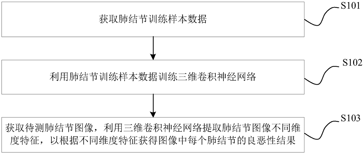
[0033] The embodiment of the present application provides a neural network-based pulmonary nodule image recognition method and device, which can effectively improve the accuracy of benign and malignant pulmonary nodule recognition results, and do not require manual judgment by doctors to improve work efficiency.

[0034] In order to enable those skilled in the art to better understand the technical solutions in the present invention, the technical solutions in the embodiments of the present invention will be clearly and completely described below in conjunction with the drawings in the embodiments of the present invention. Obviously, the described The embodiments are only some of the embodiments of the present invention, not all of them. Based on the embodiments of the present invention, all other embodiments obtained by persons of ordinary skill in the art without making creative efforts shall fall within the protection scope of the present invention.

[0035] The following w...


PUM


Abstract

Embodiments of the present invention provide a neural network based pulmonary nodule image recognition method and device. The method includes: acquiring pulmonary nodule training sample data, whereinthe pulmonary nodule training sample data includes pulmonary nodule training sample data marked with benign attributes and pulmonary nodule training sample data marked with malignant attributes; training a three-dimensional convolutional neural network by using the pulmonary nodule training sample data as input data; acquiring a pulmonary nodule image to be tested, inputting the pulmonary nodule image to be tested into the three-dimensional convolutional neural network, extracting image features of different dimensions of the pulmonary nodule image to be tested by using the three-dimensional convolutional neural network, and obtaining the benign and malignant discrimination results of each pulmonary nodule in the pulmonary nodule image according to the image features of different dimensions. The three-dimensional convolutional neural network of the embodiment of the invention can accurately distinguish the benign and malignant pulmonary nodules, effectively improve the accuracy of thebenign and malignant distinguishing results of the pulmonary nodules, and improve the working efficiency without manual judgment by the doctor.

Description

technical field [0001] The embodiments of the present application relate to the field of computer technology, and in particular to a neural network-based lung nodule image recognition method and device. Background technique [0002] In recent years, medical imaging technology has developed rapidly, improving doctors' ability to diagnose various diseases. For example, the application of computed tomography (Computed Tomography, CT) medical images can assist doctors in diagnosing whether a patient has lung cancer. In the prior art, a doctor generally judges whether a pulmonary nodule is benign or malignant according to the characteristics of the CT image by reading the CT image. With the increase in the number of patients, this manual reading method has the defects of low efficiency and low accuracy. At present, there is already a computer-aided detection method, which can obtain the approximate area of ​​​​pulmonary nodules in CT images through image processing, and then ma...

Claims


Application Information

Patent Timeline
IPC IPC(8): G06K9/62G06N3/04
CPCG06V2201/03G06N3/045G06F18/241G06F18/214
Inventor 鞠光亮
Owner SHENYANG NEUSOFT MEDICAL SYST CO LTD
Why Eureka
Try Eureka