
A CT image pulmonary nodule detection method based on improved Faster R-CNN framework

Inactive Publication Date: 2018-12-11
SOUTHEAST UNIV
View PDF0 Cites 22 Cited by
  • Summary
  • Abstract
  • Description
  • Claims
  • Application Information

AI Technical Summary

Problems solved by technology

Traditional pulmonary nodule detection methods can only detect large nodules, but for small nodules, often miss detection

Method used


Image

  • A CT image pulmonary nodule detection method based on improved Faster R-CNN framework
  • A CT image pulmonary nodule detection method based on improved Faster R-CNN framework
  • A CT image pulmonary nodule detection method based on improved Faster R-CNN framework

Examples


Embodiment Construction

[0024] The present invention will be further explained below in conjunction with the accompanying drawings and specific embodiments.

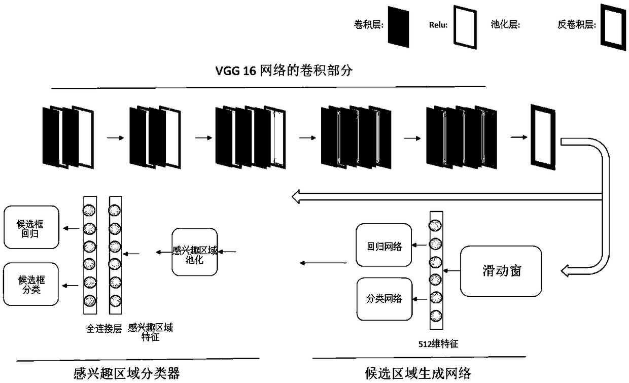
[0025] A method for detecting lung nodules in CT images based on the improved Faster R-CNN framework, comprising steps:

[0026] Step 1. Collect a CT image of the chest of a patient with pulmonary nodule symptoms, and mark the position of the pulmonary nodule as a training sample set;

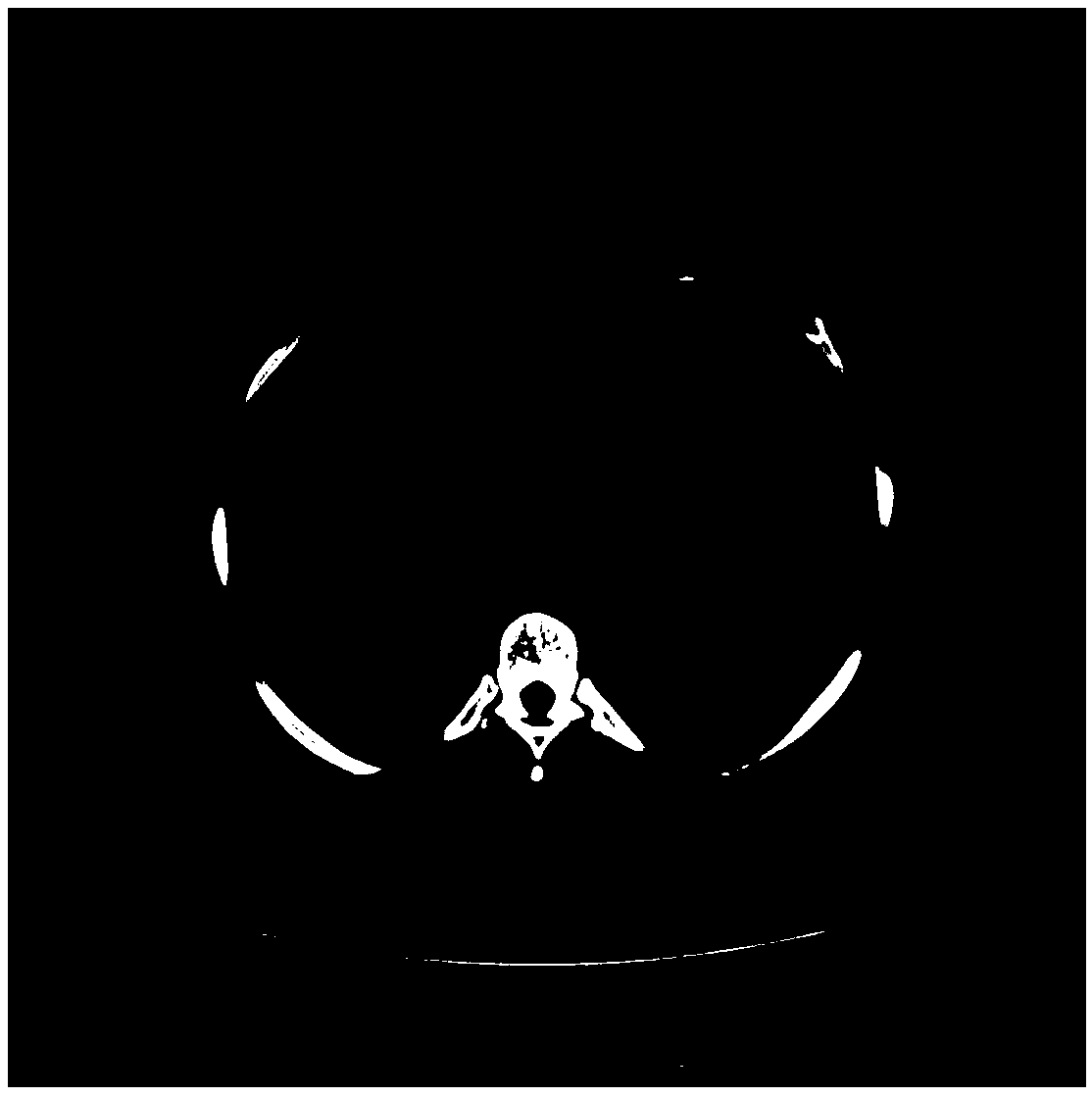
[0027] In this example, the data set published by The Lung Image Database Consortium (LIDC) is used. This data set contains 1018 cases, and each case has a corresponding experienced radiologist (usually a diagnostic information from three or four radiologists).

[0028] When processing the samples in the data set, for each patient's CT image, first interpolate the three dimensions to 1mm×1mm×1mm at the same time; since [-1200,600]HU is a more suitable range for observing the lungs, select [ The -1200,600]HU interval is mapped to [0,255] pixels; then three adja...


PUM


Abstract

The invention discloses a CT image pulmonary nodule detection method based on an improved Faster R-CNN framework. The method comprises the following steps: 1, collecting the CT images of the chest ofthe patients with pulmonary nodule symptoms, and marking the position of the pulmonary nodule as a training sample set; 2, constructing a candidate nodule detection network, training that candidate nodule detection network with a training sample set, determining network parameters, and obtaining a candidate nodule detection model; 3, constructing a false positive rejection classification network of candidate nodules, and training the false positive rejection classification network of candidate nodules with a training sample set to obtain a false positive rejection classification network modelof candidate nodules; 4, inputting a CT image to be detected into a candidate nodule detection model to obtain a position of the candidate nodule; inputting the position of the candidate nodules intoa false positive rejection classification network model of the candidate nodules, eliminating false positive rejection, and obtaining the detection result of the lung nodules. This method is more suitable for the detection of small pulmonary nodules.

Description

technical field [0001] The invention relates to a method for detecting lung nodules in CT images, in particular to a method for detecting lung nodules in CT images based on an improved Faster R-CNN framework, and belongs to the technical field of lung nodule detection. Background technique [0002] X-ray Computer Tomography (CT) technology is an imaging technology that obtains accurate and non-destructive cross-sectional attenuation information of an object by performing ray projection measurement on an object. It is one of the conventional and effective clinical medical diagnostic tools. Providing rich three-dimensional human organ tissue information for clinicians' diagnosis and prevention has become an indispensable inspection and diagnosis method in the field of medical imaging. [0003] Lung cancer has the highest mortality rate among all cancers, with a mortality rate of 13% for men and 19.5% for women. About 70% of patients are diagnosed with lung cancer at an advanc...

Claims


Application Information

Patent Timeline
IPC IPC(8): G06T7/00G06K9/62G06N3/04G06N3/08
CPCG06N3/08G06T7/0012G06T2207/10081G06T2207/30064G06N3/045G06F18/24
Inventor 陈阳葛治文蔡宁罗立民
Owner SOUTHEAST UNIV
Learn More