Myocardial quantitative magnetic resonance imaging method, equipment and storage medium

A magnetic resonance imaging and myocardial technology, applied in the field of medical imaging, can solve the problems of long scanning time, low signal-to-noise ratio, affecting the quality of T1 fitting, etc., and achieve the effect of improving spatial resolution and expanding imaging field of view.

Active Publication Date: 2021-08-10
TSINGHUA UNIV
View PDF0 Cites 0 Cited by
  • Summary
  • Abstract
  • Description
  • Claims
  • Application Information

AI Technical Summary

Problems solved by technology

Therefore, there is a problem of long scan time and mismatch between different sampling points
And, due to the part T 1 The signal-to-noise ratio of weighted sampling points is relatively low, which affects the final T 1 fit quality

Method used


Image

  • Myocardial quantitative magnetic resonance imaging method, equipment and storage medium
  • Myocardial quantitative magnetic resonance imaging method, equipment and storage medium
  • Myocardial quantitative magnetic resonance imaging method, equipment and storage medium

Examples


Embodiment Construction

[0030] In order to make the objects, technical solutions, and advantages of the present invention more apparent, an exemplary embodiment according to the present invention will be described in detail below with reference to the accompanying drawings. Obviously, the described embodiments are merely the embodiments of the present invention, rather than all embodiments of the present invention, which will be appreciated that the invention is not limited by the example embodiments described herein. Based on the embodiments described in the present invention, those skilled in the art should fall within the scope of the invention without paying creative labor.

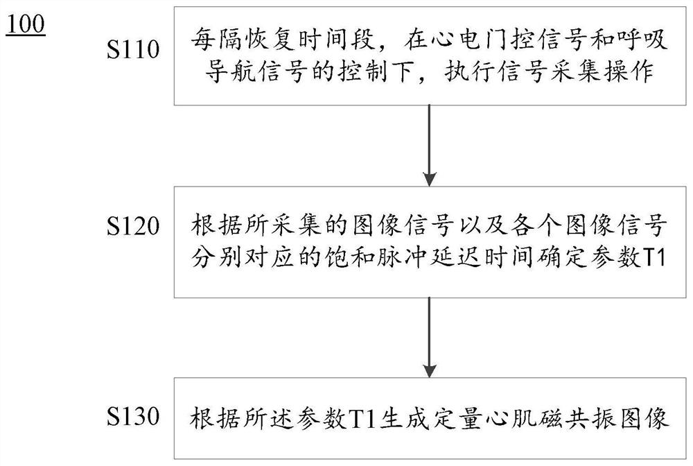
[0031] According to an embodiment of the present invention, a cardiomycological magnetic resonance imaging method is provided. This method is a 3D free breathable quantitative myocardial parameter T 1 Imaging technology. This technology adopts respiratory navigation technology to achieve compensation for respiratory motion. Base...


PUM


Abstract

The invention provides a myocardial quantitative magnetic resonance imaging method, equipment and storage medium. The method includes: performing an image signal acquisition operation under the control of the ECG gating signal and the respiratory navigation signal at intervals of the recovery period; determining the parameter T according to the acquired image signal and the delay time of the corresponding saturation pulse 1 ;according to the parameter T 1 Generate myocardial quantitative magnetic resonance images. This protocol allows the scan to be completed with the subject breathing freely, without the need for breath-holding. At the same time, it also allows to further expand the imaging field of view and improve the spatial resolution. In addition, segmented acquisitions are fully interleaved between sampling points via k‑space, enabling intrinsic registration of the original image without additional image processing in post.

Description

Technical field [0001] The present invention relates to the field of medical imaging, and more particularly to a cardium-quantitative magnetic resonance imaging method, apparatus, and storage medium. Background technique [0002] Nuclear magnetic resonance imaging techniques utilize nuclear magnetic resonance phenomena to human imaging, which is already a common medical imaging examination. [0003] Basic physical parameters of nuclear magnetic resonance T 1 (Self-ribs relaxation time, or a longitudinal relaxation time) is a time constant that describes the longitudinal magnetization vector recovery. T 1 The composition of the biological tissue is determined in the form of a structure and the magnetic field strength. At the determined magnetic field strength, different tissues have specific T 1 Numerical value. When the biological tissue changes, T 1 It will also change. So T 1 Myocardial tissue characteristics can be identified as feature parameters. [0004] Myocardial T in rec...

Claims


Application Information

Patent Timeline
Patent Type & Authority Patents(China)
IPC IPC(8): A61B5/055
CPCA61B5/0044A61B5/055A61B5/7203A61B5/7225A61B2576/023
Inventor 丁海艳郭瑞罗建文
Owner TSINGHUA UNIV
Why Eureka
Try Eureka