Dual-energy computer X-ray tomography device

A tomography and X-ray technology, applied in the field of dual-energy computer X-ray tomography devices, can solve the problems of sacrificing image quality, high cost, and unsatisfactory results.

Pending Publication Date: 2019-01-11
SHANDONG DACHENG MEDICAL TECH CO LTD
View PDF5 Cites 2 Cited by
  • Summary
  • Abstract
  • Description
  • Claims
  • Application Information

AI Technical Summary

Problems solved by technology

This method requires the addition of expensive detectors, and the spectral separation method will be inferior to other dual-energy CT implementations due to probability attenuation.
[0007] The above implementation methods either require expensive costs or sacrifice image quality in practical applications, and the results are not satisfactory.

Method used


Image

  • Dual-energy computer X-ray tomography device
  • Dual-energy computer X-ray tomography device
  • Dual-energy computer X-ray tomography device

Examples


Embodiment 1

[0067] A dual-energy computerized X-ray tomography device, including a radiation source and a detector, a collimator is arranged between the radiation source and the detector, and an imaging area and a dual-energy module are placed in sequence along the radiation direction between the collimator and the detector , The radiation source uses Varex GS-5071 bulb and Spellman X5427 high voltage generator (HVG).

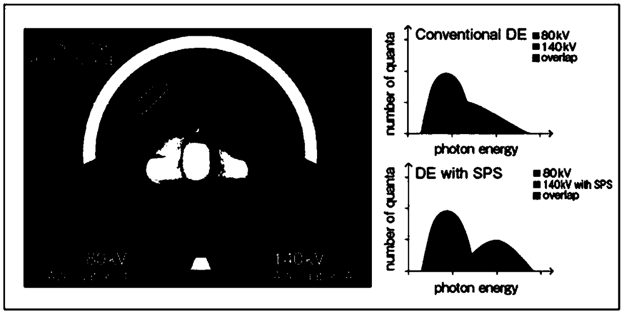
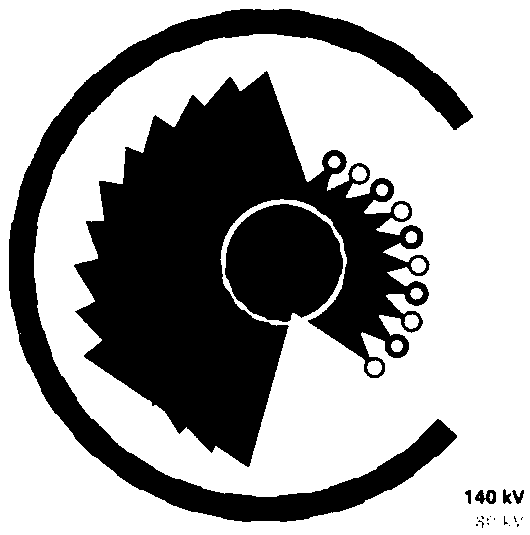
[0068] The dual-energy module includes two single-material butterfly filters made of aluminum 1100, which are used for head and body scanning respectively; two dual-energy filters are used to separate the ray beam.

[0069] The dual-energy filter includes a butterfly-shaped filter body made of aluminum 1100. The first absorption zone and the second absorption zone are arranged side by side at the bottom. The first absorption zone is made of gold and the second absorption zone is made of tin. Under the configuration of Varex GS-5071 tube and Spellman X5427 high voltage generator...

Embodiment 2

[0073] A dual-energy computerized X-ray tomography device, including a radiation source and a detector, a collimator is arranged between the radiation source and the detector, and an imaging area and a dual-energy module are placed in sequence along the radiation direction between the collimator and the detector , The radiation source uses Varex GS-5071 bulb and Spellman X5427 high voltage generator (HVG).

[0074] The dual-energy module includes two single-material butterfly filters made of aluminum 1100, which are used for head and body scanning respectively; two dual-energy filters are used to separate the beam of rays.

[0075] The dual-energy filter includes a butterfly filter body, made of aluminum 1100. The bottom is arranged side by side with a first absorption zone and a second absorption zone. The first absorption zone is made of gold and the second absorption zone is made of tin. Under the configuration of Varex GS-5071 tube and Spellman X5427 high voltage generator (HVG...

Embodiment 3

[0078] A dual-energy computerized X-ray tomography device, including a radiation source, a detector, a collimator is arranged between the radiation source and the detector, and a dual-energy module is arranged between the radiation source and the collimator, and the collimator and the detector The imaging area is set between the detectors, and the radiation source is Varex GS-5071 bulb and Spellman X5427 high voltage generator (HVG).

[0079] The dual-energy module includes two single-material butterfly filters made of aluminum 1100, which are used for head and body scanning respectively; two dual-energy filters are used to separate the ray beam.

[0080] The dual-energy filter includes a butterfly-shaped filter body made of aluminum 1100. The first absorption zone and the second absorption zone are arranged side by side at the bottom. The first absorption zone is made of gold and the second absorption zone is made of tin. Under the configuration of Varex GS-5071 tube and Spellman ...


PUM


Abstract

The invention relates to a dual-energy computer X-ray tomography device, comprising a radiation source, a filter, and a detector, wherein the filter arranged between the radiation source and the detector; the filter comprises a dual-energy module comprising at least one dual-energy filter; the dual-energy filter comprises a filter body and a filter plate, and the filter plate comprises a first absorption zone consisting of a high k-edge material and a second absorption zone consisting of a low k-edge material, arranged side by side; the k-edge value of the high k-edge material is above 60 keV;and the k-edge value of the low k-edge material is below 40kev. The dual-energy computer X-ray tomography device can realize dual-energy scanning at low cost and ensure picture quality.

Description

Technical field [0001] The invention relates to the technical field of medical imaging imaging, in particular to a dual-energy computer X-ray tomography device. Background technique [0002] In the prior art, dual-energy computed tomography (DECT) technology has multiple implementation methods: [0003] The first way, and the simplest way to achieve it, is to use the existing scanning equipment to make the tube perform two series of scans at two different voltages to obtain two completely different energy spectra. This method will not increase the equipment cost of the scanning equipment, but because the two groups need to be scanned, the time delay will reduce the speed of scanning the patient; at the same time, because the scanning duration is relatively long, the person being scanned between the two scans Physical activity can also cause image quality problems. [0004] The second way is to add a set of tubes and detectors to the rack to make the tubes work at different tube vol...

Claims


Application Information

Patent Timeline
IPC IPC(8): A61B6/03
CPCA61B6/032A61B6/482
Inventor 陈牧福瑞斯特·麦克道戈尔兰道夫·麦金利苏士刚
Owner SHANDONG DACHENG MEDICAL TECH CO LTD
Why Eureka
Try Eureka