3D reconstruction method of pathological section and device thereof

A pathological slice, 3D technology, applied in the field of 3D reconstruction method of pathological slice and its device, can solve the problem of easily missing small suspicious structures, and achieve the effect of reducing the misdiagnosis rate, accurately obtaining, and simplifying the medical process

Active Publication Date: 2019-01-11
上海全景医学影像诊断中心有限公司
View PDF4 Cites 5 Cited by
  • Summary
  • Abstract
  • Description
  • Claims
  • Application Information

AI Technical Summary

Problems solved by technology

However, it requires doctors to focus on three-dimensional imagination thinking, and it is easy to miss

Method used


Image

  • 3D reconstruction method of pathological section and device thereof
  • 3D reconstruction method of pathological section and device thereof
  • 3D reconstruction method of pathological section and device thereof

Examples


Embodiment Construction

[0034] The technical solutions of the present invention will be further described below in conjunction with the embodiments and the accompanying drawings.

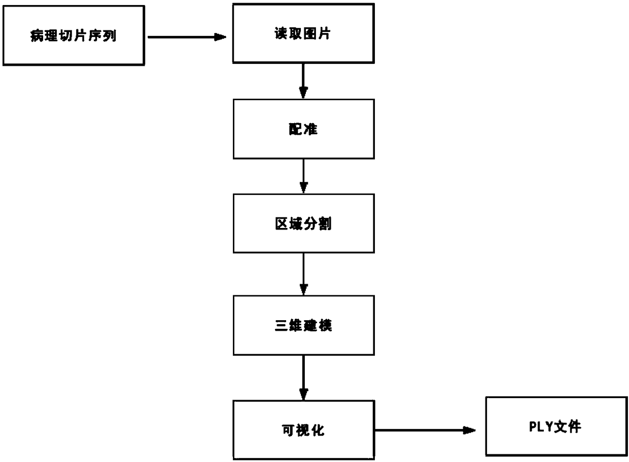
[0035] The present invention provides a kind of 3D reconstruction method of pathological section, at least comprises the following steps (referring to figure 1 The schematic flow chart of the method shown):

[0036] Step A: Acquiring digital image data corresponding to the sequence of pathological slide pictures;

[0037] Step B: Determine the position of each adjacent layer of the pathological slice according to the obtained digital image data, and generate a three-dimensional array of grid pairs;

[0038] Step C: performing data processing on the obtained three-dimensional array to obtain a binary three-dimensional array;

[0039] Step D: Carry out three-dimensional modeling according to the obtained binary three-dimensional array and save it as a three-dimensional model file;

[0040] Step E: loading the 3D model fil...


PUM


Abstract

The invention discloses a 3D reconstruction method of pathological sections, which is characterized in that the method at least comprises the following steps: step A, obtaining digital image data corresponding to a pathological slice picture sequence; step B, determining the positions of each adjacent layer of the pathological slice according to the obtained digital image data, and generating a three-dimensional array of the mesh to the adjacent layers; step C, performing data processing on the obtained three-dimensional array to obtain a binary three-dimensional array; step D, carrying out three-dimensional modeling according to the obtained binary three-dimensional array and saving as a three-dimensional model file; step E, loading the three-dimensional model file into the three-dimensional drawing software to visualize the three-dimensional structure of the pathological slice picture.

Description

technical field [0001] The present invention relates to the technical field of computer imaging, in particular to a method and device for 3D reconstruction of pathological slices applied in a medical system. Background technique [0002] With the maturity of computer technology and the development of digital information technology, earth-shaking scientific and technological progress has been made in various fields, and more and more conveniences have been provided in the fields of medical research and clinical diagnosis. The most direct applications such as digital Storage, training and teaching, and remote diagnosis are all quite mature. Based on the particularity of the medical industry, existing digital assistance systems focus more on platform provision, connection, and storage functions. The statistical software that emerges from the analysis of medical imaging materials focuses more on scientific research results, such as the foreign Slicer and the MITK platform of th...

Claims


Application Information

Patent Timeline
IPC IPC(8): G06T7/55G06T17/20
CPCG06T7/55G06T17/20G06T2200/04G06T2207/10081G06T2207/30004G06T2210/41
Inventor 王进祥吴湛
Owner 上海全景医学影像诊断中心有限公司
Why Eureka
Try Eureka