OCT system for widening fundus imaging range

An imaging range and imaging technology, applied in the field of OCT system, can solve the problems of inability to understand the condition of the human eye to be measured, and the imaging range of the fundus imaging map is small.

Pending Publication Date: 2019-02-15
ZD MEDICAL HANGZHOU CO LTD
View PDF0 Cites 1 Cited by
  • Summary
  • Abstract
  • Description
  • Claims
  • Application Information

AI Technical Summary

Problems solved by technology

[0004] In order to overcome the deficiencies in the prior art, the object of the present invention is to use an OCT system for increasing the imaging range of the fundus to solve the problem that the imaging range of the fundus imaging map is small, resulting in the inability to accurately understand the condition of the human eye under test based on the fundus tomogram

Method used


Image

  • OCT system for widening fundus imaging range
  • OCT system for widening fundus imaging range
  • OCT system for widening fundus imaging range

Examples


Embodiment Construction

[0024] Below, the present invention will be further described in conjunction with the accompanying drawings and specific implementation methods. It should be noted that, under the premise of not conflicting, the various embodiments described below or the technical features can be combined arbitrarily to form new embodiments. .

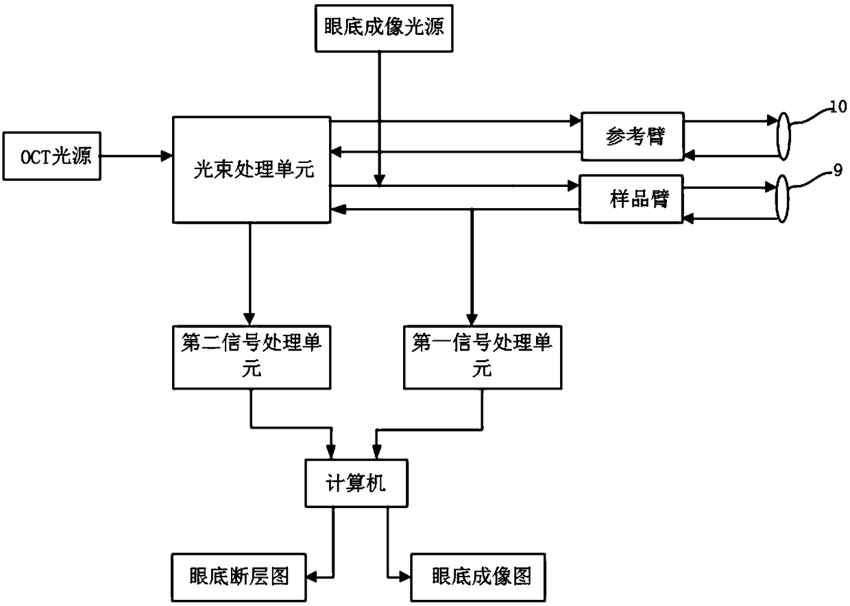
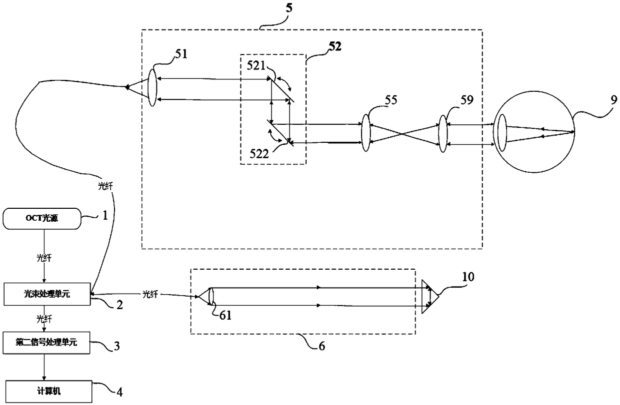
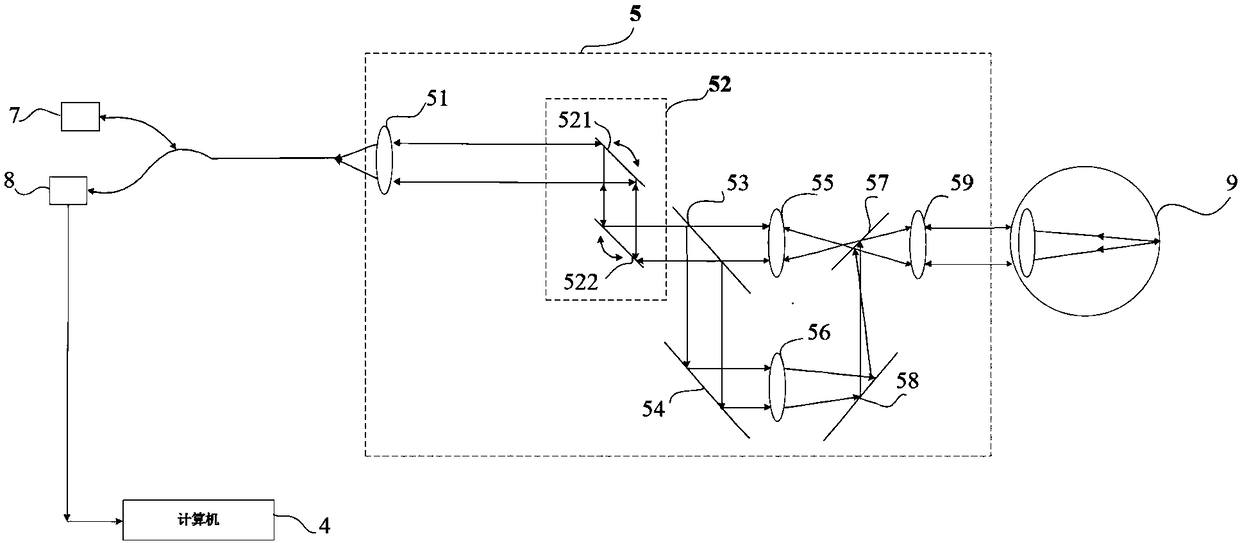
[0025] In order to solve the problem that the imaging range of the fundus imaging map is small, resulting in the inability to accurately understand the condition of the human eye under test based on the fundus tomogram, such as figure 2 , image 3 with Figure 4 As shown, the present embodiment provides an OCT system for increasing the fundus imaging range, which includes an OCT light source 1, a fundus imaging light source 7, a beam processing unit 2, a first signal processing unit 8, a second signal processing unit 3, a computer 4. The sample arm 5, the reference arm 6 and the reference mirror 10, wherein the sample arm 5 includes a vibrating mirr...


PUM


Abstract

The invention provides an OCT system for widening the fundus imaging range, and belongs to the technical field of medical imaging. The OCT system comprises a fundus imaging light source, a sample arm,a first signal processing unit and a computer; the sample arm comprises a galvanometer unit, an eye lens and a distance-lengthening scanning mirror unit; the distance-lengthening scanning mirror unitcomprises a first optical filter, a second optical filter, a first reflecting mirror, a second reflecting mirror and a first scanning lens; a first light beam emitted by the fundus imaging light source sequentially passes through the galvanometer unit, the first optical filter, the first reflecting mirror, the first scanning lens, the second reflecting mirror, the second optical filter and the eye lens to reach the detected human eyes finally, and then is reflected back the first signal processing unit from the detected human eyes along an original light path to form first data flow, and thefirst data flow forms a fundus imaging picture through a computer. The distance-lengthening scanning mirror unit is arranged between the galvanometer unit in the sample arm and the eye lens, the fundus imaging picture with the wide imaging range can be obtained, and thus the condition of the detected human eyes can be accurately understood according to a fundus fault picture.

Description

technical field [0001] The invention relates to the technical field of medical imaging, in particular to an OCT system for enlarging the fundus imaging range. Background technique [0002] OCT (Optical Coherence Tomography, optical coherence tomography) imaging is derived from ultrasonic imaging technology, which combines the characteristics of low-coherence interference and confocal microscopic measurement. By detecting the phase delay of scattered light waves, scattered light and reference light at different depths of the fundus Interference, so as to detect the reflection depth corresponding to the phase delay, and then obtain the fundus tomogram through the steps of beam scanning, signal conversion, data extraction, etc. Condition. Usually, before obtaining the fundus tomogram, it is also necessary to obtain a transverse cross-sectional view of the human eye under test, that is, a fundus imaging map, and use the fundus imaging map to determine the position where the tom...

Claims


Application Information

Patent Timeline
Patent Type & Authority Applications(China)
IPC IPC(8): A61B3/12A61B3/14
CPCA61B3/12A61B3/102A61B3/14
Inventor 胡献恩许宁莫建华
Owner ZD MEDICAL HANGZHOU CO LTD
Why Eureka
Try Eureka