A light source modulation method for confocal scanning microscope
A technology of light source modulation and microscopy, which is applied in the field of confocal scanning optical imaging instruments, can solve the problems of insufficient image quality, achieve the effect of reducing the amount of light radiation and minimizing the amount of light radiation of samples
- Summary
- Abstract
- Description
- Claims
- Application Information
AI Technical Summary
Problems solved by technology
Method used
Image
Examples
Embodiment 1
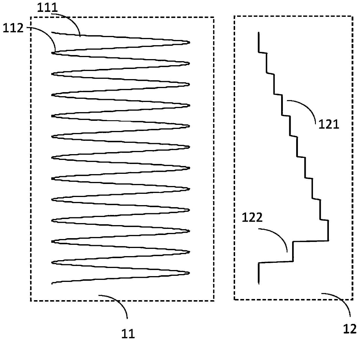
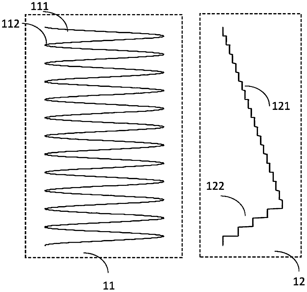
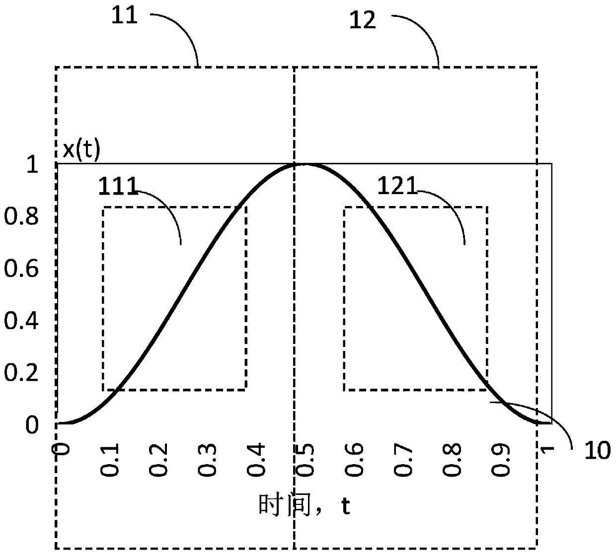
[0070] refer to Figure 2A In the image window 111 and the image window 121, the relatively linear part between the scanning window 11 and the scanning window 12 is taken. Since the sample image is severely stretched and distorted on both sides of the sample with a low speed of the fast resonant mirror, it will cause the image to be unsuitable.
[0071] In the above-mentioned embodiment 1, when a super luminescent diode (SLD, Super Luminescent Diode) is used as the imaging light source, this method is relatively easy to implement. A typical SLD usually has an opto-isolator with a built-in bandwidth of 100kHz to 200kHz, while the resonant frequency of a common fast resonant mirror is generally between 4kHz and 16kHz. Therefore, the SLD has a bandwidth of 100 kHz to 200 kHz, which is fast enough to reflect the switching frequency from the fast resonant mirror. According to the Nyquist sampling theory, it can be guaranteed that no sampling distortion will occur.
[0072] refer t...
Embodiment 2
[0079] The method adopted in this embodiment is to use a light source power modulator, and a typical application is to use an Acousto-Opto Modulator (AOM, Acousto-Opto Modulator) to modulate the light source. The modulation method starts from formula (5). In the formula (5), if the transmitting power P of the light source 0 After AOM, and then perform nonlinear modulation on AOM, get:
[0080]
[0081] Among them, k is the attenuation coefficient of the AOM, which is a constant. Substitute formula (6) into formula (5) to get:
[0082] I'(x)=ΔI(x) / Δx=k·P 0 / ω (7)
[0083] That is to say, after the power modulation of the formula (6), the radiation distribution of the light source in the sample space becomes a constant.
[0084] Simplify formula (6) further to get,
[0085]
[0086] refer to Figure 4A , which shows that simultaneous power modulation 21, 22 is performed on the output end of the light source in the forward scanning sampling window 11 and the reverse sc...
Embodiment 3
[0094] The method adopted in this embodiment is to combine the methods of Embodiment 1 and Embodiment 2 at the same time, and at the same time turn off the SLD light source outside the image window, and then superimpose a power modulator, such as an AOM, to perform power modulation on the output end of the light source.
[0095] Such as Figure 6A As shown, simultaneous power modulation 211, 221 and light source switch modulation 311, 321 are performed on the light source in the forward scanning image window 111 and reverse scanning image window 121 of the fast resonant mirror through the AOM.
[0096] Such as Figure 6B As shown, simultaneous power modulation 211 and light source switch modulation 311 are performed on the light source in the forward scanning image window 111 of the fast resonant mirror through the AOM.
[0097] Such as Figure 6C As shown, the light source is subjected to simultaneous power modulation 221 and light source switch modulation 321 through the A...
PUM
 Abstract
Description
Claims
Application Information
 - R&D
- Intellectual Property
- Life Sciences
- Materials
- Tech Scout
- Unparalleled Data Quality
- Higher Quality Content
- 60% Fewer Hallucinations
Browse by: Latest US Patents, China's latest patents, Technical Efficacy Thesaurus, Application Domain, Technology Topic, Popular Technical Reports.
© 2025 PatSnap. All rights reserved.Legal|Privacy policy|Modern Slavery Act Transparency Statement|Sitemap|About US| Contact US: help@patsnap.com


