Tissue image processing method and device, storage medium and computer equipment

A technology for organizing images and processing methods, which is applied in the field of medical image data processing, and can solve problems such as reducing diagnostic efficiency and diagnostic accuracy

Active Publication Date: 2019-03-26
SHANGHAI UNITED IMAGING HEALTHCARE
View PDF7 Cites 1 Cited by
  • Summary
  • Abstract
  • Description
  • Claims
  • Application Information

AI Technical Summary

Problems solved by technology

However, since there are many types of tissues contained in medical images, when a doctor observes one of the tissues, other types of tissues may interfere with the image o

Method used


Image

  • Tissue image processing method and device, storage medium and computer equipment
  • Tissue image processing method and device, storage medium and computer equipment
  • Tissue image processing method and device, storage medium and computer equipment

Examples


Embodiment Construction

[0035] In order to make the purpose, technical solution and advantages of the present application clearer, the present application will be further described in detail below in conjunction with the accompanying drawings and embodiments. It should be understood that the specific embodiments described here are only used to explain the present application, and are not intended to limit the present application.

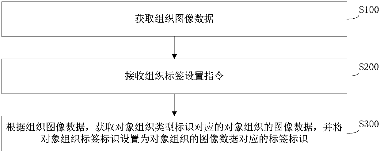
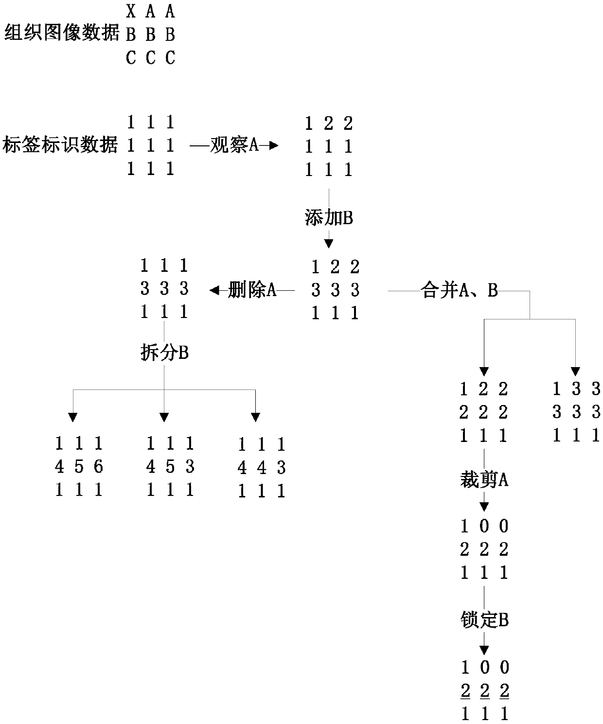
[0036] In the post-processing of medical images, it is usually targeted to observe and diagnose certain tissue structures of the human body. During the diagnosis process, multiple different tissues can be separated by processing the medical images, and the tissue images can be modified. and display. For example: for the head and neck data of CT scan, after the head and neck bone removal algorithm, the blood vessels and bone tissue can be extracted separately, and the doctor can observe and analyze the carotid artery or the skull area. The tissue image processing method pr...


PUM


Abstract

The invention relates to a tissue image processing method and device, a storage medium and computer equipment. The method includes the steps of obtaining tissue image data; receiving a tissue label setting instruction; setting label identifiers of object tissue as label identifiers corresponding to image data of the object tissue. After the tissue image data is obtained, when management operationneeds to be carried out on the tissue involved in the tissue image data, the tissue label setting instruction is received in order to set the label identifiers of the image data of the object tissue,correspondingly the label identifiers of the object tissue are changed, different label identifiers correspond to different rendering parameters, the corresponding tissue images are thus distinguishedfrom other tissue images during display, doctors can more conveniently carry out further management and observation on the tissue images to obtain diagnosis information, and therefore the diagnosis efficiency is improved. Meanwhile, other tissue can be prevented from generating image interference on the object tissue, and therefore the diagnosis accuracy is improved.

Description

technical field [0001] The present application relates to the technical field of medical image data processing, in particular to a tissue image processing method, device, storage medium and computer equipment. Background technique [0002] In the medical field, it is a widely used technology to obtain the medical data of the detected object and perform medical diagnosis based on the corresponding medical image. The medical image can well display various tissue information of the detected object, thus helping doctors to conduct diagnosis. [0003] In traditional technologies, medical images obtained from medical data often contain a variety of tissues, and doctors judge whether the tissues are normal based on the image information of the tissues to be detected in the medical images. However, since there are many types of tissues contained in medical images, when a doctor observes one of the tissues, other types of tissues may interfere with the image of the tissue, and the d...

Claims


Application Information

Patent Timeline
IPC IPC(8): G16H30/40
CPCG16H30/40
Inventor 苏刚
Owner SHANGHAI UNITED IMAGING HEALTHCARE
Why Eureka
Try Eureka