Cancer medical image data processing method, system and device and storage medium

A medical image and data processing technology, applied in the field of image processing, can solve the problems of low analysis accuracy and efficiency, easy loss of information, etc., to overcome the sensitivity to noise, improve the efficiency and accuracy of recognition and cropping

Inactive Publication Date: 2019-05-21
SOUTH CHINA UNIV OF TECH
View PDF5 Cites 19 Cited by
  • Summary
  • Abstract
  • Description
  • Claims
  • Application Information

AI Technical Summary

Problems solved by technology

[0006] When using traditional networks such as unimproved UNet for image processing, it is easy to lose information during continuous sampling
[0007] The above problems have caused the analysis accuracy and efficiency of the prior art to be at a low level

Method used


Image

  • Cancer medical image data processing method, system and device and storage medium
  • Cancer medical image data processing method, system and device and storage medium
  • Cancer medical image data processing method, system and device and storage medium

Examples


Embodiment Construction

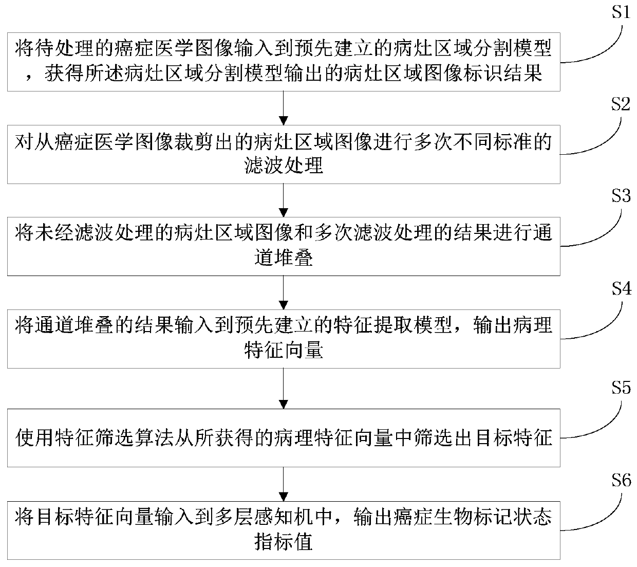
[0061] A method for processing cancer medical image data, referring to figure 1 , including the following steps:

[0062] S1. Input the cancer medical image to be processed into the pre-established lesion area segmentation model, and obtain the lesion area image identification result output by the lesion area segmentation model;

[0063] S2. Perform multiple filtering processes with different standards on the image of the lesion area cut out from the cancer medical image;

[0064] S3. Perform channel stacking on the unfiltered image of the lesion area and the result of multiple filtering processes;

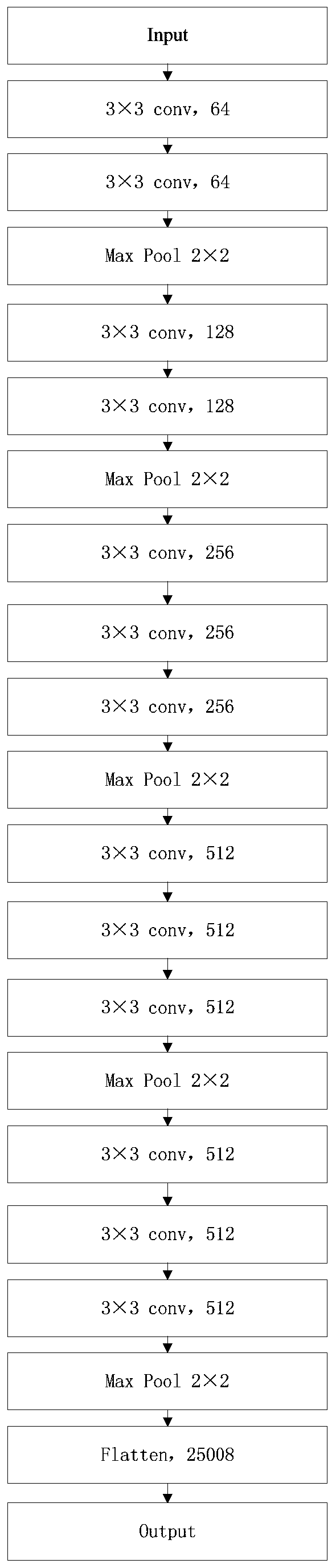
[0065] S4. Input the result of channel stacking into the pre-established feature extraction model, and obtain the pathological feature vector output by the feature extraction model; the pathological feature vector has the depth feature information of the result of channel stacking;

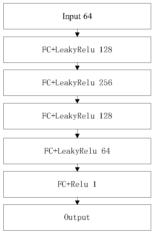
[0066] S5. Use a feature screening algorithm to screen out target features from the obtained pat...


PUM


Abstract

The invention discloses a cancer medical image data processing method, system, device and a storage medium. The method comprises the following steps: inputting a to-be-processed cancer medical image into a pre-established focus area segmentation model, obtaining a focus area image identification result output by the focus area segmentation model, filtering processing of different standards is carried out on the focus area image cut from the cancer medical image for multiple times, channel stacking is carried out, a channel stacking result is input into a pre-established feature extraction model, and a feature screening algorithm is used for screening out target features from the obtained pathological feature vectors; inputting the target characteristics into a multilayer perceptron to obtain cancer biomarker state index values output by the multilayer perceptron, and the like. According to the method, the focus area on the cancer medical image can be automatically identified, and the focus area image is cut out, so that the identification and cutting efficiency and accuracy are greatly improved. The method is widely applied to the technical field of image processing.

Description

technical field [0001] The present invention relates to the technical field of image processing, in particular to a cancer medical image data processing method, system, device and storage medium. Background technique [0002] In the field of cancer diagnosis and treatment, it is often necessary to analyze medical images such as CT and MRI of patients, locate lesions and extract features of lesions. However, the existing medical image analysis technology generally has the following problems: [0003] Before extracting the features of the lesion area, it is necessary to identify and cut out the lesion area of ​​the small target from the medical image to eliminate the interference of irrelevant information. At present, this process needs to be completed manually, and the efficiency is extremely low. Image processing, while the accuracy of cropping the lesion area is low; [0004] In the process of feature extraction, manual features are extracted using existing technologies s...

Claims


Application Information

Patent Timeline
Patent Type & Authority Applications(China)
IPC IPC(8): G06T7/00G06T7/11G06N3/04
Inventor 高英罗雄文王锦杰成昱霖
Owner SOUTH CHINA UNIV OF TECH
Why Eureka
Try Eureka