Animal brain tissue section microscopic image segmentation method

A technology for microscopic images and tissue slices, applied in image analysis, image data processing, 2D image generation, etc., to achieve the effect of ensuring accuracy

Pending Publication Date: 2019-06-07
ANHUI NORMAL UNIV
View PDF1 Cites 6 Cited by
  • Summary
  • Abstract
  • Description
  • Claims
  • Application Information

AI Technical Summary

Problems solved by technology

[0004] In view of this, the purpose of the present invention is to propose a microscopic image segmentation method for animal brain tissue sections to solve all or part of the deficiencies in the prior art

Method used


Image

  • Animal brain tissue section microscopic image segmentation method
  • Animal brain tissue section microscopic image segmentation method

Examples


Embodiment Construction

[0038] In order to make the object, technical solution and advantages of the present invention clearer, the present invention will be described in further detail below in conjunction with specific embodiments and with reference to the accompanying drawings.

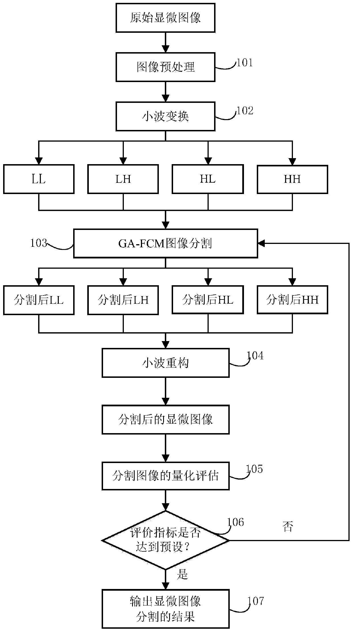
[0039] Embodiments of the present invention provide a method for segmenting microscopic images of animal brain tissue slices in order to solve all or part of the deficiencies of the current technology. The segmentation method includes the following steps:

[0040] (1) Microscopic image preprocessing;

[0041] (2) Microscopic image wavelet transform;

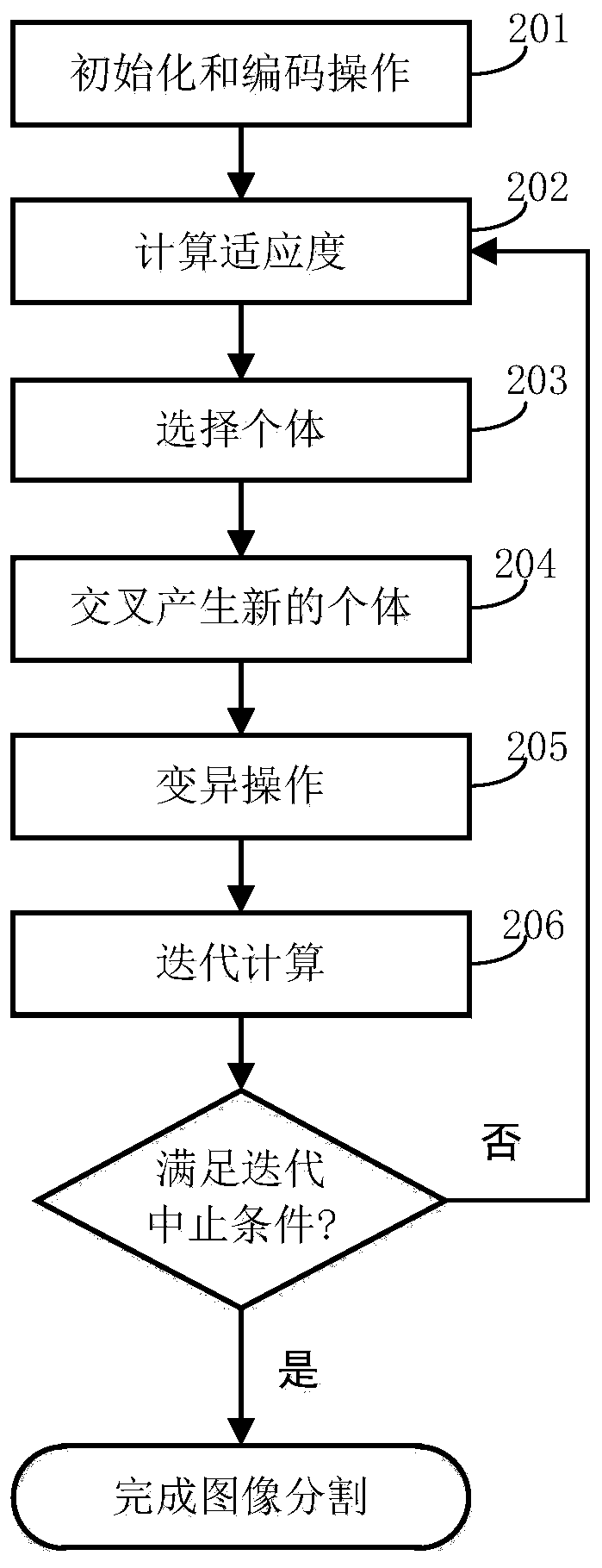
[0042] (3) GA-FCM image segmentation;

[0043] (4) Microscopic image wavelet reconstruction;

[0044] (5) Quantitative evaluation of enhanced images;

[0045] (6) Analyze whether the evaluation index reaches the preset;

[0046] (7) Output the result of microscopic image segmentation.

[0047] In order to make the purpose, technical solutions and advantages of the embod...


PUM


Abstract

The invention discloses an animal brain tissue section microscopic image segmentation method, and relates to the technical field of microscopic image processing, and the method comprises the steps: (1) carrying out the preprocessing of a microscopic image; (2) carrying out wavelet transformation on the microscopic image; (3) performing GA-FCM image segmentation; (4) reconstructing the wavelet of the microscopic image; (5) performing quantitative evaluation on the enhanced image; (6) analyzing whether the evaluation index reaches a preset index; and (7) outputting a microscopic image segmentation result: firstly, processing the microscopic image by virtue of wavelet transform to obtain high-frequency and low-frequency subspace images, and then respectively segmenting the high-frequency subspace image and the low-frequency subspace image, so that the high-frequency component and the low-frequency component of the microscopic image are effectively segmented, and GA-FCM is adopted to realize the effective segmentation of the microscopic image, and evaluates the segmentation effect by means of the quantitative index of objective evaluation, thereby ensuring the segmentation accuracy ofthe microscopic image, and meeting the requirement of subsequent three-dimensional reconstruction of the brain tissue structure.

Description

technical field [0001] The invention relates to the technical field of microscopic image processing, in particular to a method for segmenting microscopic images of animal brain tissue slices. Background technique [0002] With the rapid development of computer technology and the continuous updating of biomicroscopic imaging technology and equipment, the establishment of digital virtual human, animal and three-dimensional visualization models of their organs based on anatomical structures has become a research hotspot in various countries. Studying the structure of animal brain tissue and constructing a three-dimensional visualization model of animal brain tissue can more clearly grasp its three-dimensional structure, which is of great significance for related research and teaching of zoology. [0003] In order to realize the three-dimensional reconstruction of the animal brain tissue structure, it is necessary to use a microscope to observe the structure of the animal brain ...

Claims


Application Information

Patent Timeline
Patent Type & Authority Applications(China)
IPC IPC(8): G06T7/168G06T11/00G06K9/62G06N3/12
Inventor 刘再群李蓓柯成炎
Owner ANHUI NORMAL UNIV
Why Eureka
Try Eureka