X-ray apparatus having a composite field of view

一种X射线、视场的技术,应用在辐射诊断用隔膜、放射诊断临床应用、用于放射诊断的仪器等方向,能够解决放射科低效率、X射线剂量增加等问题

Pending Publication Date: 2019-07-30
KONINKLIJKE PHILIPS NV
View PDF9 Cites 4 Cited by
  • Summary
  • Abstract
  • Description
  • Claims
  • Application Information

AI Technical Summary

Problems solved by technology

Such events are undesirable, lead to inefficiencies in the radiology department, and lead to an increase in the X-ray dose absorbed by the patient

Method used


Image

  • X-ray apparatus having a composite field of view
  • X-ray apparatus having a composite field of view
  • X-ray apparatus having a composite field of view

Examples


Embodiment Construction

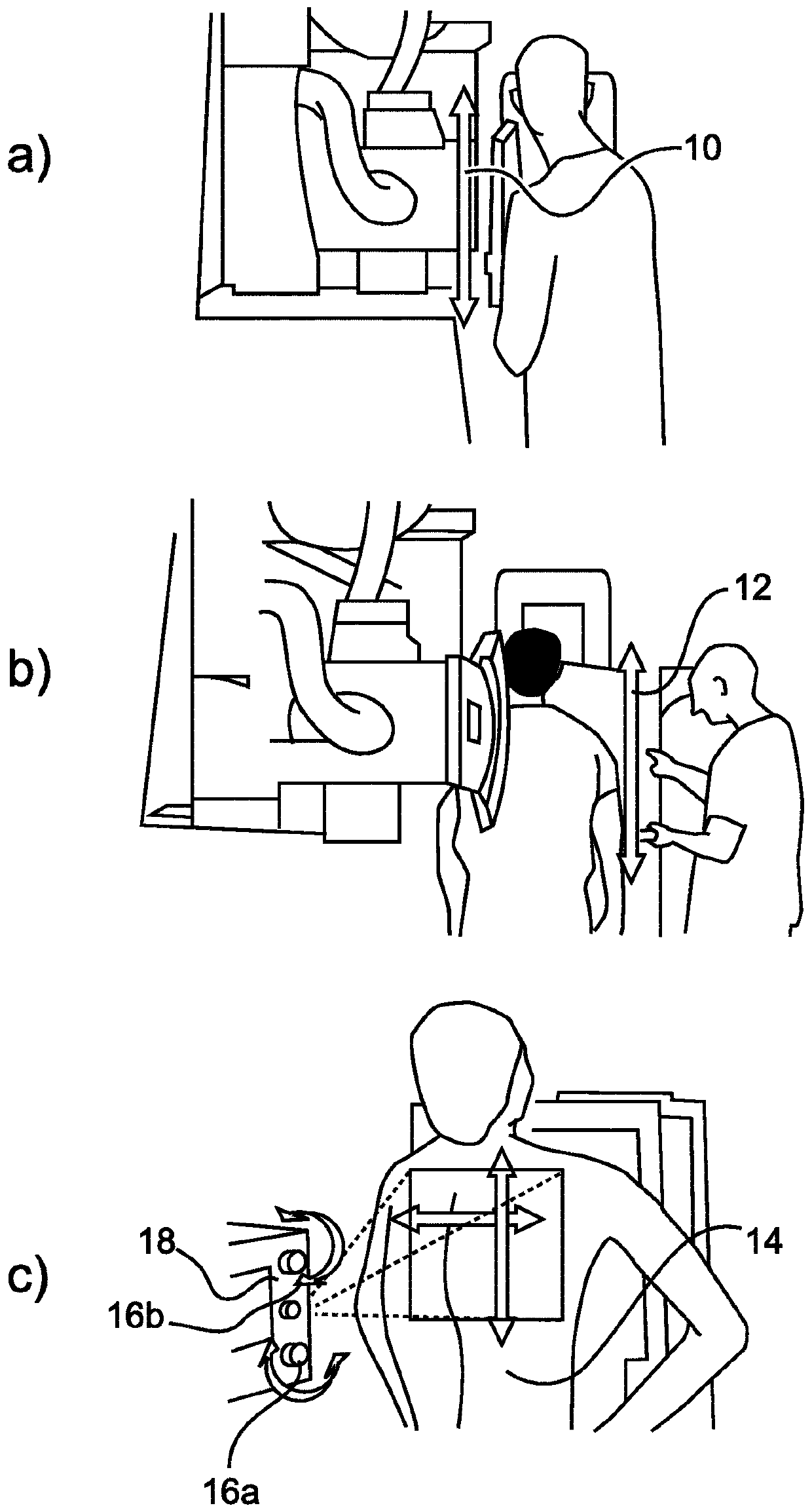
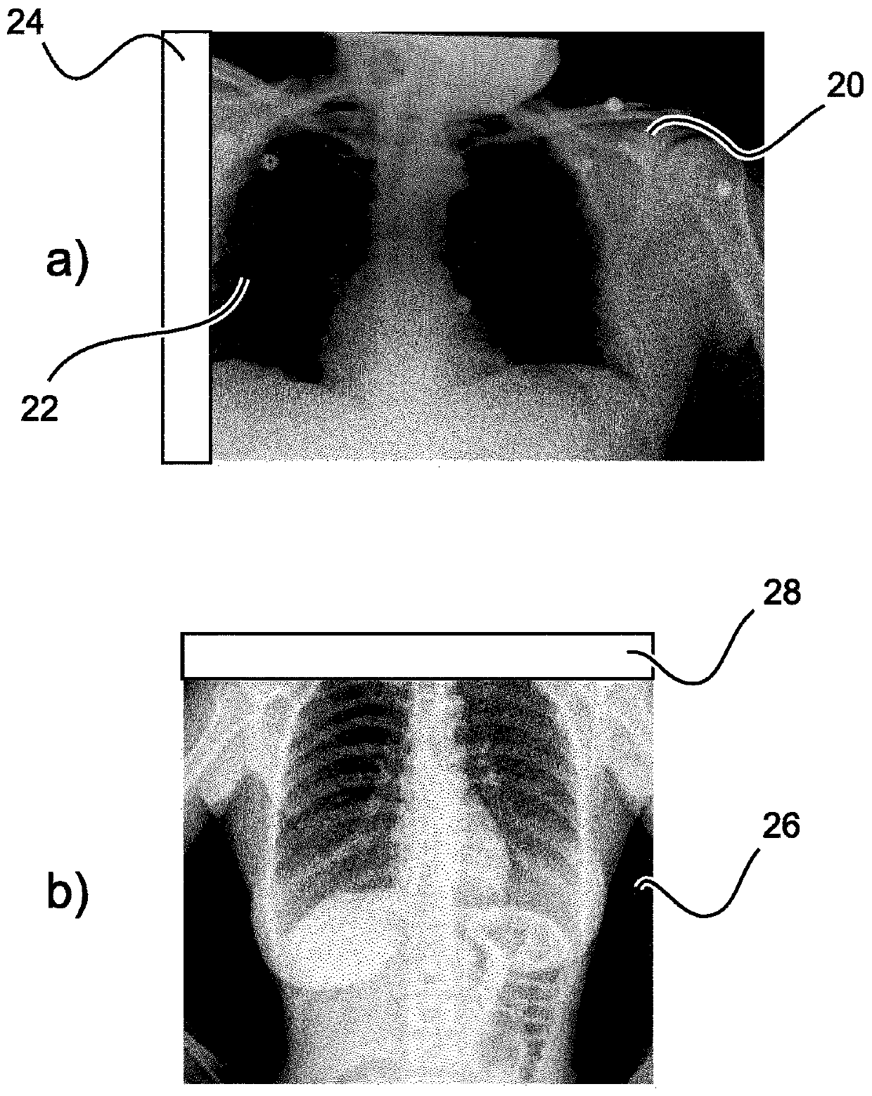
[0065] Chest radiography is the most commonly performed clinical imaging exam and plays an important role in the detection and diagnosis of many diseases of the chest anatomy. Image quality depends on a wide range of specific individual factors, such as the inclusion of appropriate anatomical structures within the field of view, the contrast of structures of interest relative to background signal, and aspects of the positioning of the patient's chest relative to the X-ray acquisition equipment.

[0066] The task of setting the field of view (FOV) for exposure is typically performed by a radiology technologist. The patient is initially positioned in the region of interest in front of the x-ray detector. Visible light irradiated from within the tube head of the X-ray equipment and matched to the field of the X-ray radiation pattern is then used to create a field of view on the patient's body. First the height of the tube head can be changed, and then the height of the "grid" co...


PUM


Abstract

Medical radiography requires specialist control of radiography equipment to achieve good imaging results. Typical errors that can occur consist of an inappropriate field of view being accidentally applied. This results in a "cropping effect" in which portions of the region of interest of a patient which would be of clinical use are omitted from the image. Conventionally, the only solution is to re-take the entire image with a more appropriate (and inevitably larger) field of view selected. This is undesirable, because it might require recall of the patient from another location, and the patient will be subject to two exposures, thus undesirably increasing their X-ray dosage. The present application proposes to use an anatomical atlas to analyse an X-ray image output from an initial exposure, in particular to assess whether significant anatomical elements are missing from the image. If elements are missing, the field of view of the X-ray imager is recalibrated, using collimation or pan / tilt adjustments, for example. A subsequent X-ray image is obtained, and combined with the initial image, to provide an output image. Because only a small area of the region of interest may subsequently need to be exposed, a smaller additional dose results.

Description

technical field [0001] The present invention generally relates to a device for obtaining an X-ray image with a composite field of view. An X-ray imaging system, a method for obtaining an X-ray image with a composite field of view, a computer program element and a computer readable medium are also discussed. Background technique [0002] Configuration of the field of view (FOV) for radiography exposures, such as pulmonary radiography, is a non-trivial task that requires the attention of a trained radiographer operating the X-ray imager. An incorrectly set field of view prior to exposure means that a subsequent full X-ray image has to be taken, since important anatomical details can be missing from the first image. Patients may even need to be recalled for a second visit. Such events are undesirable, lead to inefficiencies in the radiology department, and lead to an increase in the X-ray dose absorbed by the patient. [0003] US 2015 / 0228071 A1 discusses an apparatus and me...

Claims


Application Information

Patent Timeline
Patent Type & Authority Applications(China)
IPC IPC(8): A61B6/00A61B6/06
CPCA61B6/06A61B6/50A61B6/5241A61B6/545G06T7/0014G06T2207/10116G06T2207/20221G06T2207/30061G06T2207/30168
Inventor S·扬D·贝斯特罗夫J·冯贝格
Owner KONINKLIJKE PHILIPS NV
Why Eureka
Try Eureka